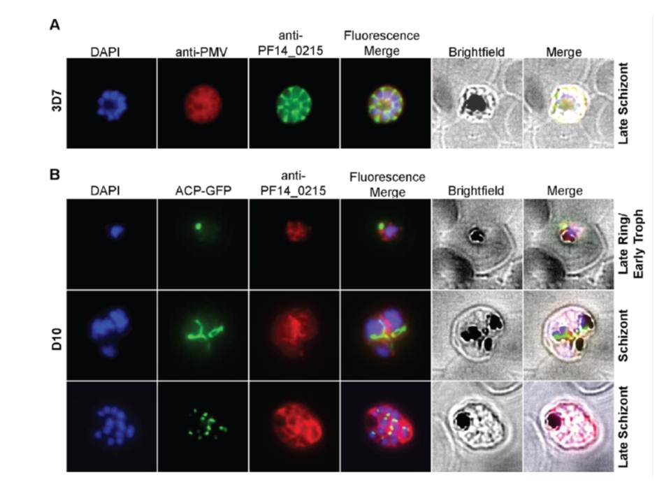
Cellular localization of HRD1. (A) HRD1 and plasmepsin V (PMV), an ER membrane marker, co-localize in P. falciparum. (B) At various stages of the parasite life cycle, HRD1 was co-stained with the nuclei (DAPI) and the apicoplast (ACP-GFP) in the P. falciparum strain D10. HRD1 was found within reticular structures outside the nuclear regions at the trophozoite and schizont stages of the parasite, similar to the physical attributes of the ER. In the late schizont stage HRD1 proteins reside within globular structures surrounding each budding merozoite in a pattern typical of the ER. These observations support that HRD1-E3 ubiquitin ligase resides in the ER membrane, consistent with a putative role in the ERAD pathway.
Chung DW, Ponts N, Prudhomme J, Rodrigues EM, Le Roch KG. Characterization of the Ubiquitylating Components of the Human Malaria Parasite's Protein Degradation Pathway. PLoS One. 2012;7(8):e43477.
Other associated proteins
| PFID | Formal Annotation |
|---|---|
| PF3D7_1323500 | PEXEL protease plasmepsin V |
| PF3D7_1422500 | ERAD-associated E3 ubiquitin-protein ligase HRD1 |