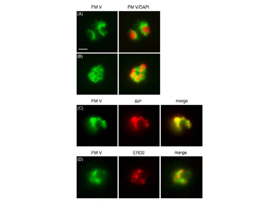
Localization of plasmepsin V (PM V). Indirect immunofluorescence assays of PM V (green) in trophozoites (A) and a ~6N schizont (B) in formaldehyde-fixed blood smears. Nuclei were visualized with DAPI (pseudocolored red). (C, D) Colocalization of PM V and the ER protein BiP (C) or the Golgi marker ERD2 (D) in trophozoites. Bar, 2 mm. In trophozoites and schizonts, PM V was concentrated around the nucleus with lower levels dispersed in the surrounding cytoplasm (Fig. 5A and B). These patterns of fluorescence suggest that PM V resides in the endoplasmic reticulum (ER), which in P. falciparum has been shown to include the nuclear envelope.
Klemba M, Goldberg DE. Characterization of plasmepsin V, a membrane-bound aspartic protease homolog in the endoplasmic reticulum of Plasmodium falciparum. Mol Biochem Parasitol. 2005 143:183-191. Copyright Elsevier 2010.
Other associated proteins
| PFID | Formal Annotation |
|---|---|
| PF3D7_0917900 | PfHsp70-2 |
| PF3D7_1323500 | PEXEL protease plasmepsin V |