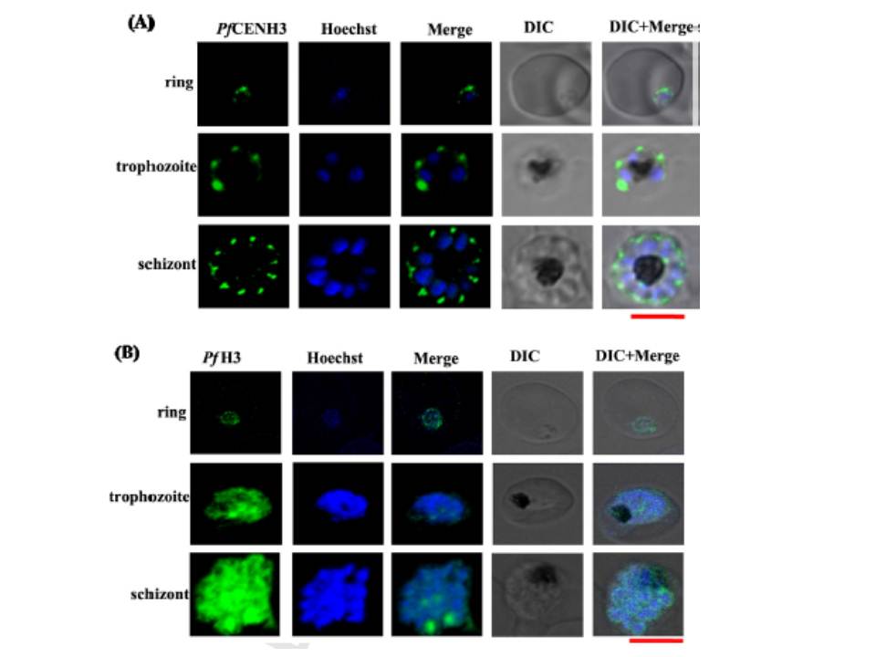
PfCENH3 is expressed at the perinuclear regions while PfH3 localizes to nucleus. (A, B) The confocal images for immuno-fluorescence assays represent the subcellular localization pattern of PfCENH3 and PfH3 (green) with respect to the Hoechst stained nucleus (blue) at ring, trophozoite and schizont stages of P. falciparum respectively. While PfH3 localizes throughout the nuclear mass, PfCENH3 localizes towards perinuclear regions at various intraerythrocytic stages. At ring stage, PfCENH3 localizes as a single cluster per nucleus. At trophozoite and schizont stages it localizes as discrete spots on each nucleus. Scale bar = 5 mm.
Verma G, Surolia N. Plasmodium falciparum CENH3 is able to functionally complement Cse4p and its, C-terminus is essential for centromere function. Mol Biochem Parasitol. 2013 Dec 6. [Epub ahead of print]
Other associated proteins
| PFID | Formal Annotation |
|---|---|
| PF3D7_1333700 | histone H3-like centromeric protein CSE4 |