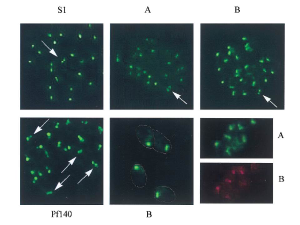
Immunofluorescence assays performed with antisera against fragments of the shared domain (S1), fragments of the unique regions, anti-PfRBP2-Ha (A) and anti-PfRBP2-Hb (B), and the rhoptry antigen Pf-140, on air-dried, acetone-fixed, P. falciparum-infected erythrocytes. The lower center panel (B) is at a higher magnification (33,750; others are 31,250), with the outline of the merozoites marked. The lower right shows costaining of single merozoites with rabbit anti-A serum (A) and rat anti-B serum (B). Each of the PfRBP2-H antisera, whether raised against fragments of the shared domain, anti-S1 and anti-S3, or against the unique regions, anti-A and anti-B, yielded a punctate staining pattern, generally with a single dot of fluorescence, although occasionally two foci were visible in a single merozoite (marked by arrows Upper). This double-dot pattern is reminiscent of proteins such as the Pf140 protein, which are localized to the paired rhoptry organelles. Higher magnification (Lower Center) reveals that the staining is concentrated at the apical end of the merozoites.
Rayner JC, Galinski MR, Ingravallo P, Barnwell JW. Two Plasmodium falciparum genes express merozoite proteins that are related to Plasmodium vivax and Plasmodium yoelii adhesive proteins involved in host cell selection and invasion. Proc Natl Acad Sci U S A. 2000 97:9648-53. Copyright 2009 National Academy of Sciences, U.S.A.
Other associated proteins
| PFID | Formal Annotation |
|---|---|
| PF3D7_1335400 | reticulocyte binding protein 2 homologue a |