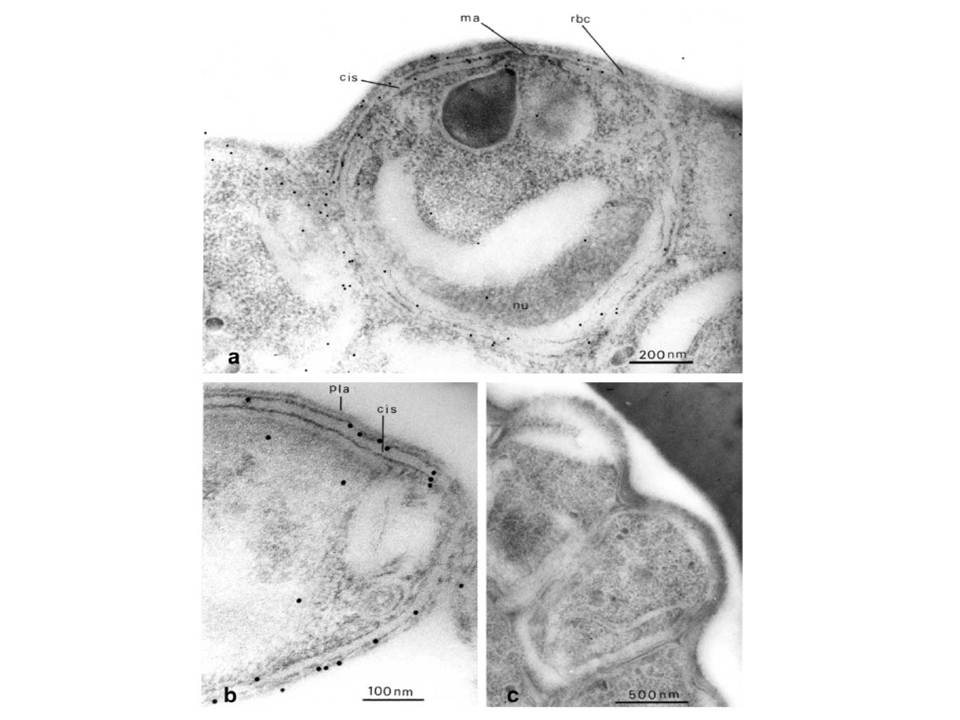
Immuno-electron microscopy, showing distribution of Pf-myo1 in the malaria parasite. (a) Immunogold-labelled merozoites before their release from the red cell. The parasite was labelled with anti-Pf-myo1 and second antibody, conjugated with 10 nm gold particles. The myosin is located around the periphery of the merozoite. In the centre the merozoite plasma membrane overlies the apical prominence (ma) close to the large pearshaped secretory rhoptry, and elsewhere in the parasite a flat cisterna (cis) underlies the plasma membrane. The curved nucleus (nu) is visible at the base of the merozoite. Note absence of label from the flat tip of the apical prominence, and the greater frequency of labelling around the apical prominence than elsewhere. (b) Longitudinal/oblique section through the apical end of a free merozoite. Labelling is associated with the cortical cytoplasm between the plasma membrane (pla) and the outer membrane of the subplasmalemmal cisterna (cis). (c) Control section through a group of mature merozoites at the periphery of a late-stage schizont, incubated with preimmune serum before gold labelling. No significant labelling can be observed.
Adapted with permission from Pinder JC, Fowler RE, Dluzewski AR, Bannister LH, Lavin FM, Mitchell GH, Wilson RJ, Gratzer WB. Actomyosin motor in the merozoite of the malaria parasite, Plasmodium falciparum: implications for red cell invasion. J Cell Sci. 1998 111:1831-9.