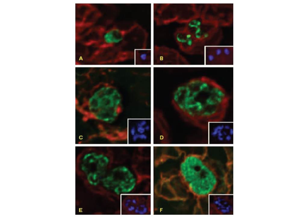
Deconvolution immunofluorescence microscopy of PfNT1 in P. falciparum-infected red blood cells. Parasites were examined by deconvolution microscopy using illumination at 546 nm to visualize PfNT1 (panels A–D), PfHT1 (panel E), and P. falciparum protein phosphatase 2C (panel F) conjugated to the FITC-conjugated anti-rabbit secondary antibody or at 488 nm to visualize Band 3 complexed with the Texas Red-conjugated anti-mouse secondary antibody (all panels). Panel A depicts a ring stage parasite, panel B shows two trophozoites, and schizonts are exhibited in panels C–F. Each panel is accompanied by an inset displaying nuclear staining with Hoechst dye.
Rager N, Mamoun CB, Carter NS, Goldberg DE, Ullman B. Localization of the Plasmodium falciparum PfNT1 nucleoside transporter to the parasite plasma membrane. J Biol Chem. 2001 276:41095-9.
Other associated proteins
| PFID | Formal Annotation |
|---|---|
| PF3D7_0204700 | Sugar transporter |