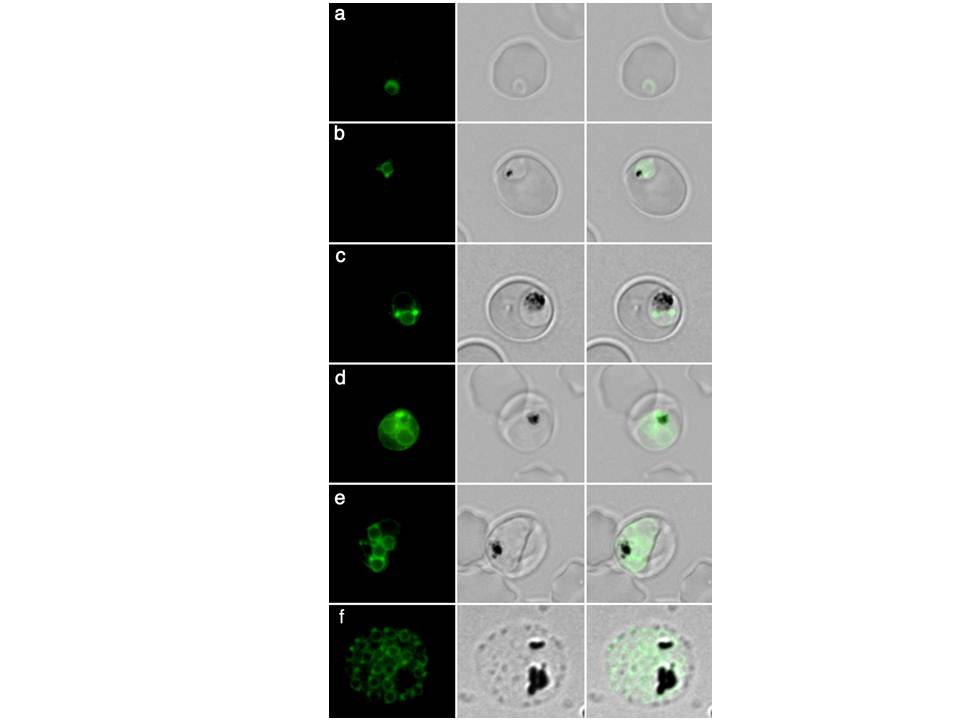
Live transgenic parasites expressing PfERD2-GFP were imaged by fluorescence microscopy. In early ring stages (8 hours post invasion, a) a weak crescent-shaped fluorescence is observed. At 16-24 hours post invasion two protrusions of intense PfERD2-GFP fluorescence are connected with weaker perinuclear staining (b-c). By 32-40 hours post invasion ongoing schizogony is observed. Nuclear division is indicated by additional, multiple perinuclear distributions of PfERD2-GFP (d-e). At 48 hours post invasion the late schizont stage is reached. Note that every forming merozoite displays one focused PfERD2-GFP signal (f).
Struck NS, de Souza Dias S, Langer C, Marti M, Pearce JA, Cowman AF, Gilberger TW. Re-defining the Golgi complex in Plasmodium falciparum using the novel Golgi marker PfGRASP. J Cell Sci. 2005 118:5603-13