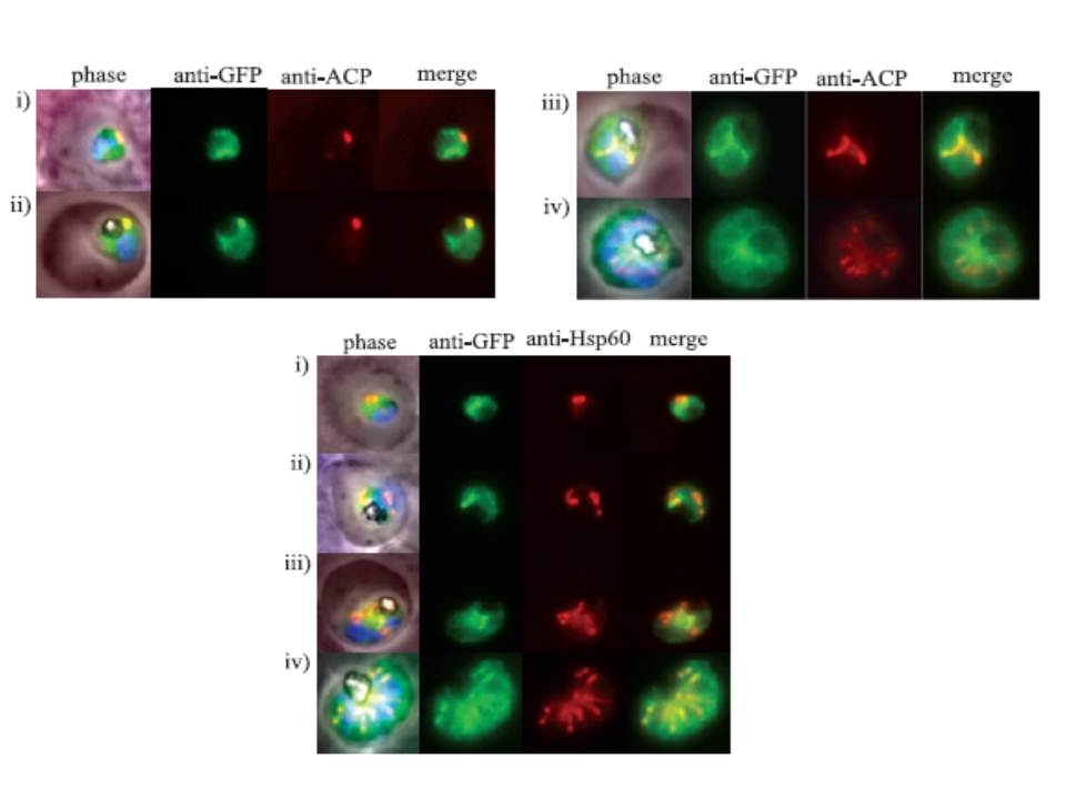
Upper panels: The intense fluorescent focus of FLN-GFP appears to partially overlap with that of the apicoplast. Clone MP3 parasites were fixed with 4% paraformaldehyde/0.0075% glutaraldehyde and stained with rabbit anti-ACP antibody, a marker for the apicoplast lumen (red) and goat anti-GFP antibody (green). Cells were observed by epifluorescence microscopy. (i–iv) Representative images of cells at ring, trophozoite, schizont and segmenter stages respectively.
LOwer panel: FLN–mitochondrion association. Clone MP3 was examined by IFA and epifluorescence microscopy using rabbit anti-Hsp60 (red) and goat anti-GFP (green) antibodies. (i–iv) Representative images of cells at ring, trophozoite, schizont and segmenter stages respectively.
Ponpuak M, Klemba M, Park M, Gluzman IY, Lamppa GK, Goldberg DE. A role for falcilysin in transit peptide degradation in the Plasmodium falciparum apicoplast. Mol Microbiol. 2007 63:314-34
Other associated proteins
| PFID | Formal Annotation |
|---|---|
| PF3D7_0208500 | acyl carrier protein |
| PF3D7_1360800 | falcilysin |