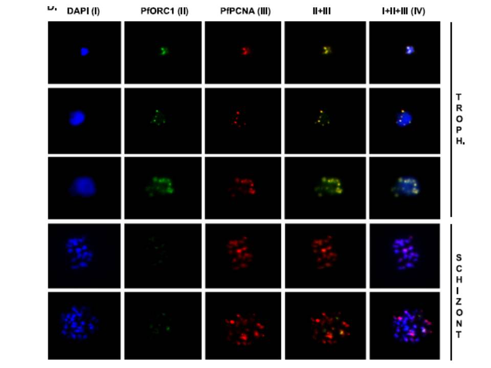
Colocalization of PfPCNA1 and PfORC1 during the replicating-trophozoite stage. A glass slide containing a parasite smear from the replicating-trophozoite stage or schizont stage was treated for immunofluoresence studies as described in Materials and Methods using both anti-PfORC1 and anti-PfPCNA1 antibodies. Distinct colocalization of PfORC1 and PfPCNA1 foci can be detected during the replicating-trophozoite stage (Troph.; rows 1 to 3). Although the expression of PfPCNA can be detected at the multinucleated late schizont stage (rows 4 and 5), the expression of PfORC1 is barely visible as the protein is degraded at the late stage, as described earlier. DAPI (4’,6-diamidino-2-phenylindole) (I) shows the nuclei. Column IV represents merged panels I, II, and III.
Gupta A, Mehra P, Deshmukh A, Dar A, Mitra P, Roy N, Dhar SK. Functional dissection of the catalytic carboxyl-terminal domain of origin recognition complex subunit 1 (PfORC1) of the human malaria parasite Plasmodium falciparum. Eukaryot Cell. 2009 8:1341-51.
Other associated proteins
| PFID | Formal Annotation |
|---|---|
| PF3D7_1361900 | proliferating cell nuclear antigen 1 |