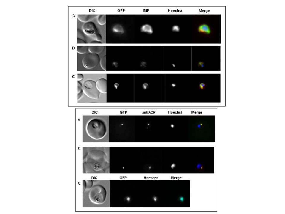
Upper panel: Subcellular localization of PfSel1, PfSel2, and PfSel4 in P. falciparum using GFP-fusion proteins. Shown are erythrocytes infected with trophozoite stage parasites. A. In the different Sel genes the UGA codon was replaced with different codons. PfSel1 (Sec->Cys), B. PfSel2 (Sec->Cys), and C. PfSel4 (Sec->Cys). Anti-BiP and Hoechst 33258 were used for co-localization with ER and nucleus, respectively. All gene products localized to the ER.
Lower panel: Subcellular localization of PfSel3 in P. falciparum using GFP-fusion proteins and different PfSel3 mutants. Shown are erythrocytes infected with trophozoite stage parasites. A. PfSel3 (Sec->Cys), B. PfSel3 (Sec->Trp), C. PfSel3-UGA. Anti-ACP and Hoechst 33258 were used for co-localization with apicoplast and nucleus, respectively. PfSel3/GFP-fusion protein, which contains its original UGA codon, localized to the nucleus. All other proteins where the UGA codon has been replaced with different codons, localized to the apicoplast.
Roeseler A, Prieto JH, Iozef R, Hecker B, Schirmer H, Kuelzer S, Przyborski J, Rahlfs S, Becker K. Insight into the selenoproteome of the malaria parasite Plasmodium falciparum. Antioxid Redox Signal. 2012 Jan 9. [Epub ahead of print]
Other associated proteins
| PFID | Formal Annotation |
|---|---|
| PF3D7_0931200 | selenoprotein |
| PF3D7_1403600 | selenoprotein |
| PF3D7_1427200 | selenoprotein |