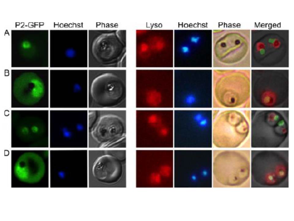
Loss of food vacuolar integrity upon fosmidomycin and wortmannin treatment Confocal fluorescence microscopic localization of a plasmepsin-II-GFP (PMII736 GFP) construct (left panels) or live fluorescence imaging of Lysotracker Red737 stained (right panels) malaria parasites. Control parasites (A) are compared to parasites treated for 24h with either fosmidomycin (+ FSM), fosmidomycin plus geranylgeraniol (+ FSM + GG-ol), or wortmannin (+ wort). Images are representative of at least three independent biological experiments. PMII-GFP was localized to the FV, which is distinguished microscopically in phase images by the presence of dark hemozoin pigment. Following fosmidomycin treatment, PMII-GFP was no longer discretely localized within the FV. Instead, PMII-GFP was dispersed throughout the parasite cytoplasm, consistent with the physical disruption of the FV that was visualized by electron microscopy.
Howe R, Kelly M, Jimah J, Hodge D, Odom AR. Isoprenoid biosynthesis inhibition disrupts Rab5 localization and food vacuolar integrity in Plasmodium falciparum. Eukaryot Cell. 2012 12(2):215-23.