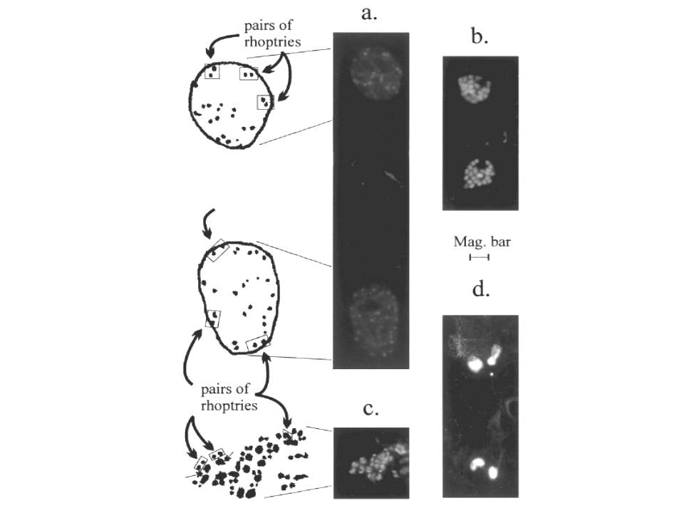
Localization of RAP-1 products in schizonts, merozoites, and rings by IFA. Fixed thin blood smears prepared from synchronized parasites were stained with the indicated primary antibody. Panel (a), schizonts stained with antibody 1E8 reveal many examples of paired organellar antibody localization indicating that the Pr86 epitope is present in the rhoptries. In the artist’s reproduction of IFA images in this panel, six of the many pairs of rhoptries are boxed. Panel (b), merozoites in segmenter-stage schizonts stained with antibody 1E8. Individual nuclei are weakly stained, and there are few, if any, examples of punctate rhoptry staining in intracellular merozoites at this late stage of schizogony. The failure of 1E8 to stain rhoptry antigen in the forming merozoites contrasts with the punctate staining of merozoites by other anti-RAP-1 antibodies (compare panel (b) with (c)). Panel (c), merozoites released from infected RBC and then stained with the anti-RAP-1 mAb 10-8G continue to exhibit a distinct paired spotty or punctate immunofluorescence pattern. Three of the many pairs of rhoptries are boxed in the reproduction to the left of panel (c), and two of the associated stained nuclei are indicated with a double-headed arrow. Panel (d), the anti-RAP-1 mAb 2D9 brightly stains diffusely distributed antigen in ring stage parasites. Panel (a) has twice the magnification of panels (b–d). Magnification bar: panel (a), 2 mm; panels (b–d), 4 mm.
Howard RF, Narum DL, Blackman M, Thurman J. Analysis of the processing of Plasmodium falciparum rhoptry-associated protein 1 and localization of Pr86 to schizont rhoptries and p67 to free merozoites. Mol Biochem Parasitol. 1998 ;92:111-22.