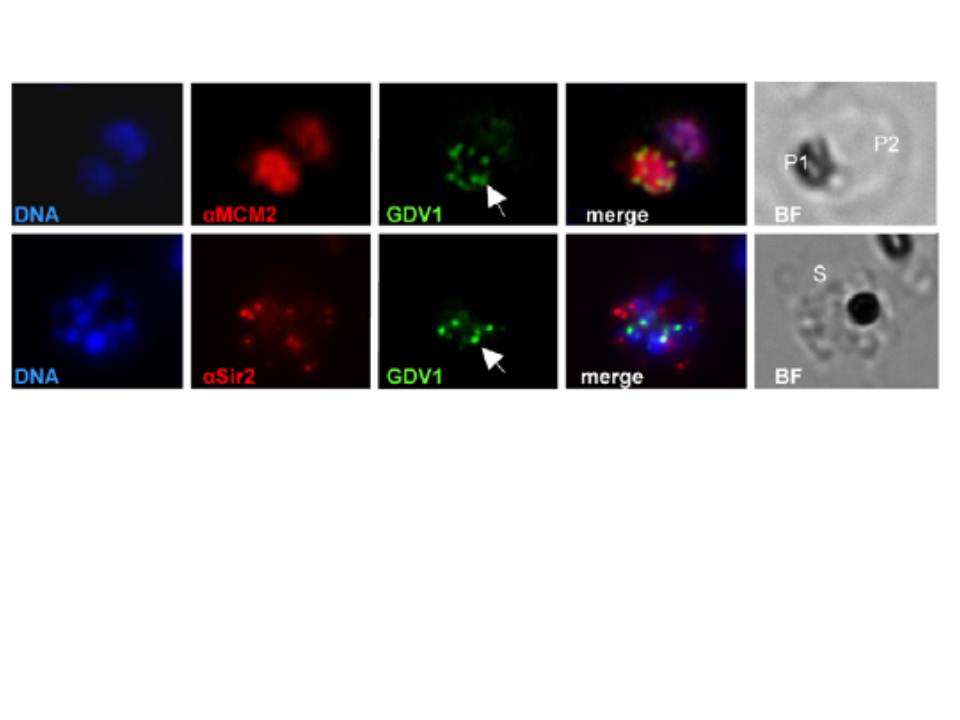
Subcellular localization of PfGDV1 (this gene product is expressed in gametocytes and is not included in MPMP). Parasites transformed with GFP- PfGDV1 were stained with DAPI (DNA stain) and the indicated anti-sera, and then examined by fluorescence microscopy (Zeiss Axiovert 200, 1000xmagnification). Images are shown of the DAPI stain (DNA), GFP-tagged PfGDV1 epifluorescence (GDV1), and antibodies specific for the indicated proteins. The corresponding merged and bright field (BF) images are included on the right. PfGDV1 expression is indicated with an arrow; locations of parasites in the BF image are indicated with a P for parasite or S for schizont. Colocalization of PfGDV1 with nuclear proteins. A doubly infected erythrocyte (Upper) with one parasite in the plane of the image (P1) and the other below (P2). Both P1 and P2 are positive for GDV1 and aMCM2. A schizont (S) expressing GDV1 stained with aSir2. MCM2 localized to the nucleus, and PfSir2 to the nuclear periphery.
Eksi S, Morahan BJ, Haile Y, Furuya T, Jiang H, Ali O, Xu H, Kiattibutr K, Suri A, Czesny B, Adeyemo A, Myers TG, Sattabongkot J, Su XZ, Williamson KC. Plasmodium falciparum gametocyte development 1 (Pfgdv1) and gametocytogenesis early gene identification and commitment to sexual development. PLoS Pathog. 2012 8(10):e1002964.
Other associated proteins
| PFID | Formal Annotation |
|---|---|
| PF3D7_1328800 | transcriptional regulatory protein sir2a |