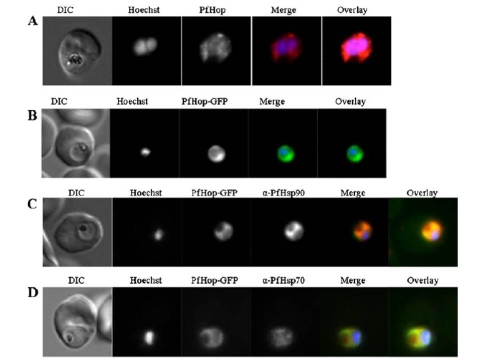
Localisation and expression of PfHop-GFP by parasites in human red blood cells. a Localisation of PfHop in P. falciparum cells; panels show a DIC image, nuclear stain (Hoechst), distribution of PfHop, merge and overlay. b Panels show a DIC image, nuclear stain (Hoechst), distribution of PfHop-GFP, merge and overlay. c Panels show a DIC image, nucleus (Hoechst), distribution of PfHop-GFP, distribution of PfHsp90, merge and overlay for PfHsp90–PfHop-GFP co-localisation. d Panels show DIC image, stain (Hoechst), distribution of PfHop-GFP, distribution of PfHsp70, merge and overlay. PfHop displayed a similar cytosolic localisation profile to PfHsp90 (b), suggesting that the two proteins may associate. Although, PfHop-GFP and PfHsp70 exhibited overlapping cytosolic co-localisation signals, the PfHsp90–PfHop-GFP co-localisation signal was more uniform than that for PfHsp70–PfHop-GFP (b, c). It is possible that PfHop associates more closely with PfHsp90 than with PfHsp70.
Gitau GW, Mandal P, Blatch GL, Przyborski J, Shonhai A. Characterisation of the Plasmodium falciparum Hsp70-Hsp90 organising protein (PfHop). Cell Stress Chaperones. 2011 17(2):191-202.
Other associated proteins
| PFID | Formal Annotation |
|---|---|
| PF3D7_0708400 | heat shock protein 90 |
| PF3D7_0818900 | PfHsp70-1 |