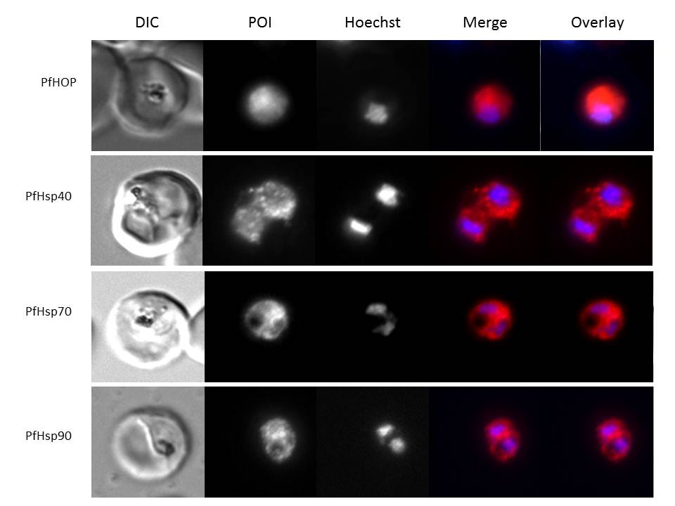
Immunofluorescence staining to detect Hsp40, HSP70, HSP90 and HOP was conducted on trophozoite stage P. falciparum-infected erythrocytes. Panels show from left to right a DIC image, distribution of protein of interest (POI), nuclear stain (Hoechst), merge and overlay (localization relative to the parasite nucleus and phase-contrast image). All proteins showed cytosolic localization.
Botha M, Chiang AN, Needham PG, Stephens LL, Hoppe HC, Külzer S, Przyborski JM, Lingelbach K, Wipf P, Brodsky JL, Shonhai A, Blatch GL. Plasmodium falciparum encodes a single cytosolic type I Hsp40 that functionally interacts with Hsp70 and is upregulated by heat shock. Cell Stress Chaperones. 2011 16(4):389-401. Pictures were kindly provided by Jude Przyborski.
PubMed Article: Plasmodium falciparum encodes a single cytosolic type I Hsp40 that functionally interacts with Hsp70 and is upregulated by heat shock
Other associated proteins
| PFID | Formal Annotation |
|---|---|
| PF3D7_0818900 | PfHsp70-1 |
| PF3D7_1434300 | Hsp70/Hsp90 organizing protein |
| PF3D7_1437900 | HSP40, subfamily A |