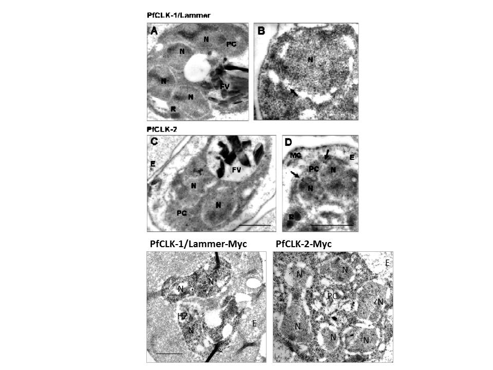
Upper two rows: Ultrastructural localization of PfCLKs in the parasite blood stages. Post-embedding immunoelectron microscopy labelling was performed using mouse antisera against PfCLK-1 and PfCLK-2, respectively, in combination with secondary gold-conjugated antibody. A: PfCLK-1/Lammer is predominantly present in the nucleus of a blood stage schizont. B: A distinct labelling of PfCLK-1 in the passage between nucleus and cytoplasm (arrow) was detected in another schizont. C: PfCLK-2 is expressed in the nucleus as well as the cytoplasm of a late trophozoite. D: PfCLK-2 labelling was particularly detected in the passage between nucleus and cytoplasm (arrows) of a schizont. E, erythrocyte; FV, food vacuole; MC, Maurer´s cleft; N, nucleus; PC, parasite cytoplasm; R, rhoptry. Bar, 0.5 μm. Lower row: Expression of Myc-tagged PfCLKs in blood stages. Post-embedding immunoelectron microscopy labelling was performed using anti-Myc antibody in combination with secondary gold-conjugated antibody. PfCLK-1/Lammer-Myc is predominantly present in the nucleus of a trophozoite. PfCLK-2-Myc can be detected in the nucleus and the cytoplasm of a schizont. E, erythrocyte; HZ, hemozoin; N, nucleus; PC, parasite cytoplasm. Bar, 0.5 μm.
Agarwal S, Kern S, Halbert J, Przyborski JM, Baumeister S, Dandekar T, Doerig C, Pradel G. Two nucleus-localized CDK-like kinases with crucial roles for malaria parasite erythrocytic replication are involved in phosphorylation of splicing factor. J Cell Biochem. 2011 112:1295-310.
Other associated proteins
| PFID | Formal Annotation |
|---|---|
| PF3D7_1445400 | protein serine/threonine kinase-1 |