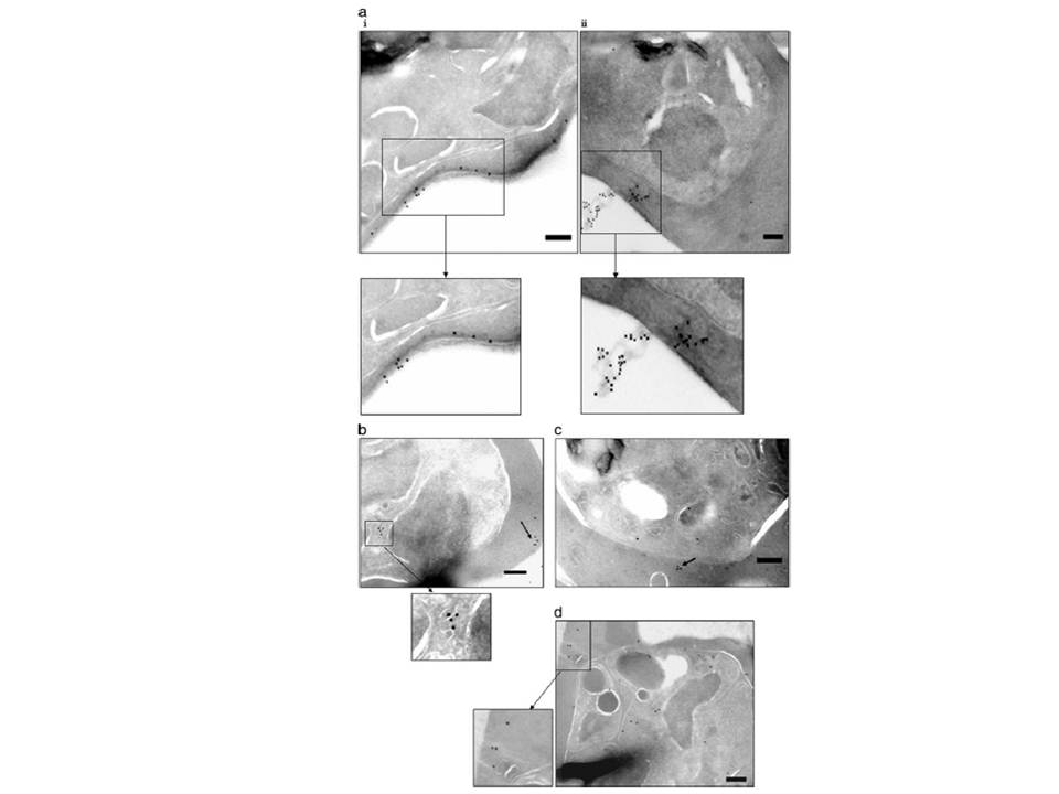
Ultrathin sections of P. falciparum-infected erythrocytes (at late trophozoite/schizont stages) were labeled with specific sera (against the four selected proteins) and gold-conjugated secondary antibody. Localization is depicted as black dots (of gold particles) for PfSEL1 (PFB0190c) - (a, i and ii), PfSEL2 = Hrd3 (b), PfEK (c), and PfEP (d). Enlarged panels and arrows show detailed images of the intracellular staining pattern. Scale bar, 250 nm. PfSEL1 showed a secreted/ extra-cellular staining pattern, localizing in the iRBC cytosol close to the erythrocyte plasma membrane. The gold particle staining showed the protein coating the surface of the iRBC and also being released out in the extra-cellular milieu. PfSEL2 (seen as dots coating vesicle-like structures) and PfEK also showed localization close to iRBC plasma membrane while being exported out of the infected red cells (b and c). PfEP localized in the extensive membranous network of the parasite from where it seemed to be trafficked to the iRBC cytosol and plasma membrane (d).
Singh M, Mukherjee P, Narayanasamy K, Arora R, Sen SD, Gupta S, Natarajan K, Malhotra P. Proteome analysis of Plasmodium falciparum extracellular secretory antigens at asexual blood stages reveals a cohort of proteins with possible roles in immune modulation and signaling. Mol Cell Proteomics. 2009 8(9):2102-18.
Other associated proteins
| PFID | Formal Annotation |
|---|---|
| PF3D7_1113100 | protein tyrosine phosphatase |
| PF3D7_1448400 | ubiquitin-protein ligase, putative |