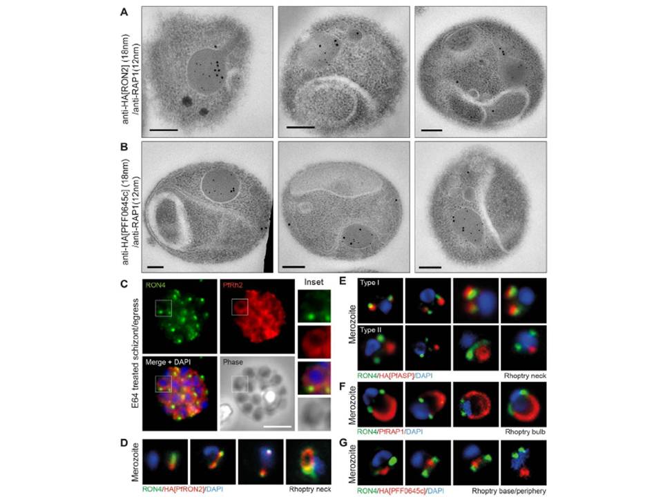
Spatial localisation of different rhoptry proteins before and during merozoite invasion. (A) IEM of free PfRON2-HA merozoites (pre-invasion) dual labeled with immunogold anti-HA (18 nm) and rhoptry bulb marker RAP1 (12 nm). Scale bar = 0.2 mm. (B) IEM of free PFF0645c-HA merozoites (pre-invasion) dual labeled with immunogold anti-HA (18 nm) and rhoptry bulb marker RAP1 (12 nm). Scale bar = 0.2 mm. (C) Widefield IFA of E64-treated schizonts (to prevent egress labeled with anti-PfRh2, anti-PfRON4 and DAPI. Scale bar = 5 mm. (D–G) Independent replicate imaging of merozoites from (D) PfRON2-HA, (E) PfASP-HA (two classes of distribution seen), (F) RAP1 and (G) PFF0645c-HA mid-way through invasion colabeled with anti-PfRON4 and DAPI. RON2-HA is located anterior to the bulb marker RAP1. PFF0645c had a more posterior or contiguous localisation to RAP1, frequently located towards the peripheral regions of the rhoptry.
Zuccala ES, Gout AM, Dekiwadia C, Marapana DS, Angrisano F, Turnbull L, Riglar DT, Rogers KL, Whitchurch CB, Ralph SA, Speed TP, Baum J. Subcompartmentalisation of Proteins in the Rhoptries Correlates with Ordered Events of Erythrocyte Invasion by the Blood Stage Malaria Parasite. PLoS One. 2012;7(9):e46160.
Other associated proteins
| PFID | Formal Annotation |
|---|---|
| PF3D7_0405900 | apical sushi protein |
| PF3D7_1116000 | rhoptry neck protein 4 |
| PF3D7_1410400 | rhoptry-associated protein 1 |
| PF3D7_1452000 | rhoptry neck protein 2 |