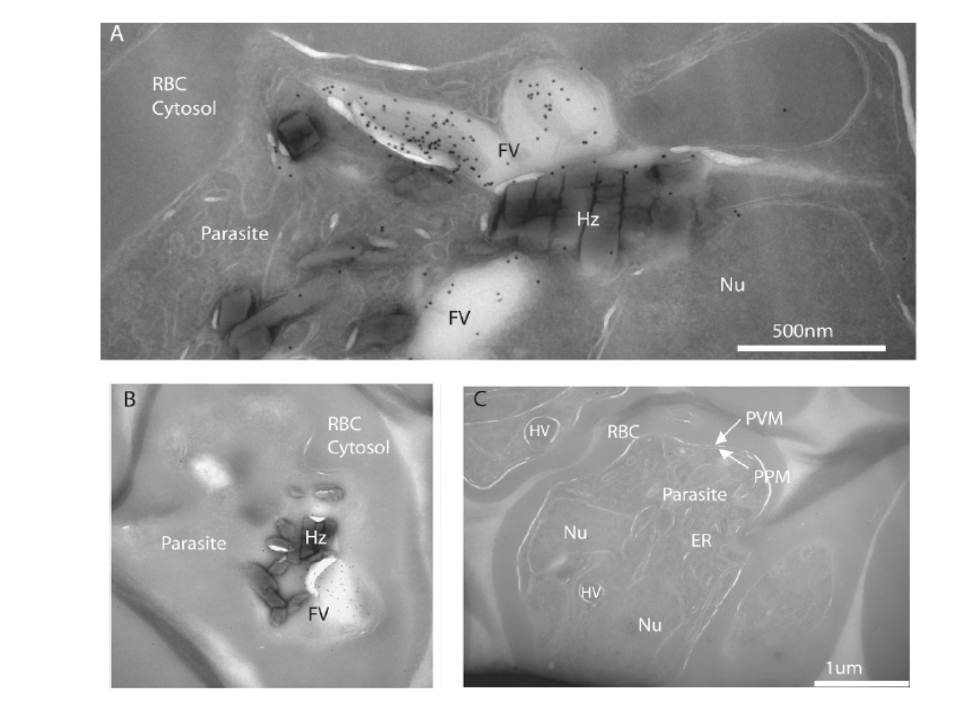
FCP localizes to the lumen of the FV. A, cryo-immunoelectron micrograph showing a close-up of a folded FV with immunogold-stained FCP in the lumen of the parasite FV in a late trophozoite. Various structures are indicated as host red blood cell cytosol (RBC Cytosol), parasite body (parasite), parasite nucleus (Nu), FV, and hemozoin (Hz). B shows a more complete parasite with a cross-section through the FV indicating a lack of immunogold staining outside the FV. C, three parasites from a different region of the same ultrathin section that was immunogold-stained for anti-FCP in A and B. Note the lack of concentrated gold particles on all visible parasite structures including ER, parasite plasma membrane (PPM), parasitophorous vacuolar membrane (PVM), and hemoglobin containing vesicles (HV).
McIntosh MT, Vaid A, Hosgood HD, Vijay J, Bhattacharya A, Sahani MH, Baevova P, Joiner KA, Sharma P. Traffic to the malaria parasite food vacuole: a novel pathway involving a phosphatidylinositol 3-phosphate-binding protein. J Biol Chem. 2007 282:11499-508.