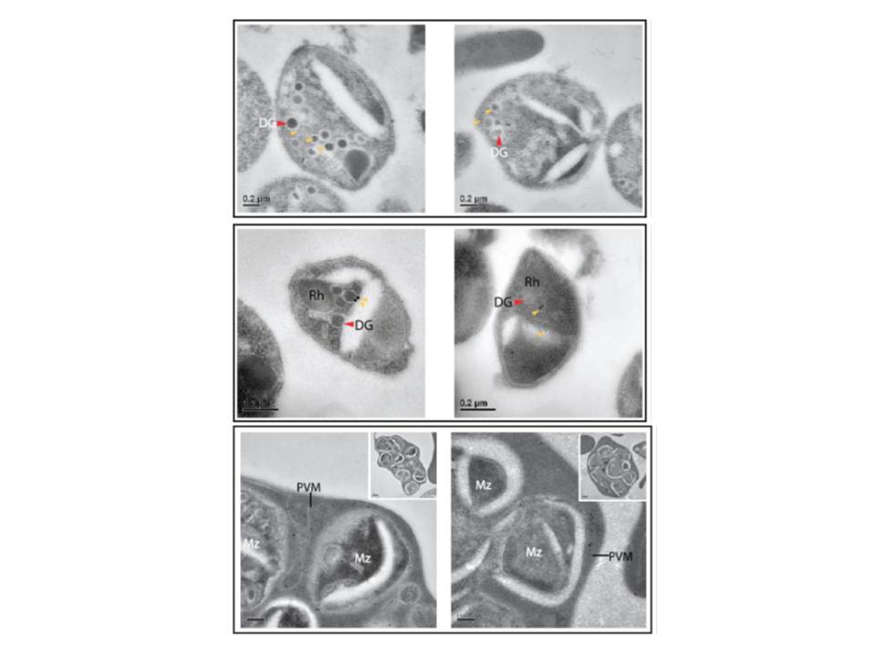
Two upper rows: PTEX components are apically localised in merozoite dense granules. Immuno-electron microscopy of isolated D10 merozoites labeled with anti-EXP2 antibodies and (D) isolated PTEX150-HA merozoites labeled with anti-HA antibodies indicate that immunogold labels (yellow triangles) localize to dense granules (DG), and not the rhoptries (Rh). Lower row: Immuno-electron microscopy of unfiltered D10 merozoites within the erythrocyte clearly resolves the PVM from the merozoite (Mz) plasma membranes. High magnification of anti-EXP2 antibody labeling localized EXP2 to the PVM (with low magnification of the entire unfiltered cell provided in the inset), indicating that it is not a plasma membrane protein and that it remains on the PVM until the point of parasite egress from the erythrocyte.
Bullen HE, Charnaud SC, Kalanon M, Riglar DT, Dekiwadia C, Kangwanrangsan N, Torii M, Tsuboi T, Baum J, Ralph SA, Cowman AF, de Koning-Ward TF, Crabb BS, Gilson PR. Biosynthesis, localisation and macromolecular arrangement of the Plasmodium falciparum translocon of exported proteins; PTEX. J Biol Chem. 2012 287(11):7871-84.