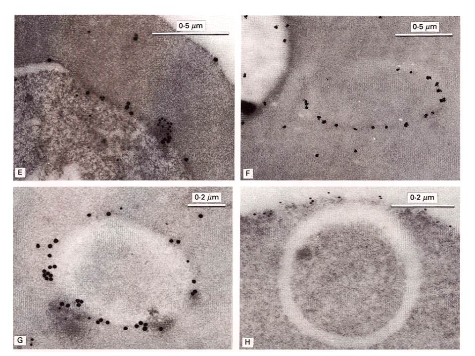
Immunoelectronmicroscopical localization of exp-2. Labelling with McAb7.7 alone (F) was followed by silver enhancement. In double-labelling studies (E, G), localization of PcAb5.1 is shown by larger, 20 nm particles; localization of McAb7.7 by 5 nm particles. (E) Co-localization of McAb7.7 and PcAb5.1 at the surface of an early schizont. Note association with a mass of dense material (lower right). (F) Localization of McAb7.7 to a membranous cleft in the erythrocyte cytoplasm (right) and low labelling of the parasite cytoplasm (left). (G) Co-localization of McAb7.7 and PcAb5.1 to a vesicular structure in the cytoplasm of an infected erythrocyte, double-labelled. (H) Plasma membrane of the RBC and intraerythrocytic membranous cleft, showing restriction of band 3 protein to the red cell surface.
Johnson D, Günther K, Ansorge I, Benting J, Kent A, Bannister L, Ridley R, Lingelbach K. Characterization of membrane proteins exported from Plasmodium falciparum into the host erythrocyte. Parasitology. 109 ( Pt 1):1-9. Copyright Cambridge University Press.