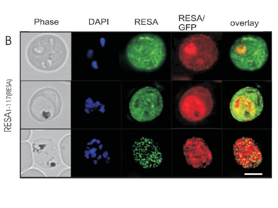
Immunofluorescence microscopy analysis of endogenous RESA and RESA1-117-GFP in transfected parasitized erythrocytes. Erythrocytes infected with RESA1-117-GFPRESA transfectants at the ring stage (top panels), trophozoite stage (middle panels), or schizont stage (bottom panels) were fixed with methanol (which destroys the intrinsic GFP fluorescence) and labeled with rabbit anti-GFP antiserum followed by an Alexa Fluor 568-conjugated anti-rabbit IgG (red fluorescence) and anti-RESA MAb, followed by fluorescein isothiocyanate-conjugated anti-mouse IgG (green fluorescence). Phase-contrast images are shown in the left panels. Overlays of the green and red fluorescence channels are shown in the right panels. Bar, 5 mm. In early-ring stage parasites, the anti-RESA antibody revealed some endogenous RESA associated with the PV which appeared to be relocated to the erythrocyte cytosol as the parasite matured. In schizont stage parasites, the antibody against endogenous RESA revealed a punctate fluorescence pattern that is consistent with labeling of dense granules in individual merozoites.
Rug M, Wickham ME, Foley M, Cowman AF, Tilley L. Correct promoter control is needed for trafficking of the ring-infected erythrocyte surface antigen to the host cytosol in transfected malaria parasites. Infect Immun. 2004 72:6095-105.