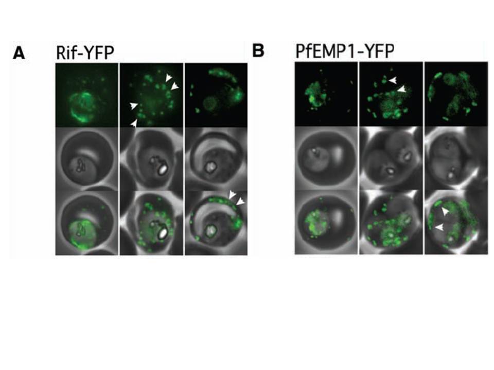
Export of Rifin surface antigens and of the major P. falciparum virulence factor, PfEMP1, depends on a functional Pexel motif. (A) A Rifin-YFP fusion including the Pexel motif is exported into the erythrocyte. Arrowheads indicate the localization of the reporter to punctate structures reminiscent of Maurer’s clefts (left panel, center) and later to the surface. (B) Export of PfEMP1-YFP also depends on a functional Pexel motif. As for the Rifin-YFP fusion, the reporter transiently localizes to structures similar to Maurer’s clefts, followed by a rim fluorescence suggestive of surface localization. Representative cells expressing the exported reporter are shown in ring stage (left), early (center), and late trophozoites (right).
Marti M, Good RT, Rug M, Knuepfer E, Cowman AF. Targeting malaria virulence and remodeling proteins to the host erythrocyte. Science. 2004 306(5703):1930-3. Copyright 2010.
Other associated proteins
| PFID | Formal Annotation |
|---|---|
| PF3D7_1240600 | erythrocyte membrane protein 1, PfEMP1 |