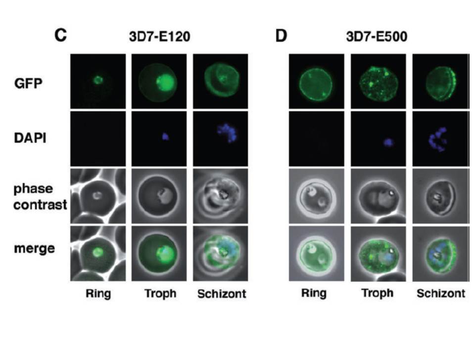
Detection of live GFP fluorescence in the PfEMP3-GFP P. falciparum transgenic lines. 3D7-E120 (C) and 3D7-E500 (D). Shown for each transgenic line is a representative image of a ring stage-infected RBC, a trophozoite stage-infected RBC and a schizont stage-infected RBC. The first row in each panel depicts the native GFP fluorescence, the second row the nucleus stained with DAPI, the third row the phase contrast image and the forth row the overlays of all three images. parasites expressing E120-GFP also generally showed an even distribution although there was some apparent concentration at the periphery of the infected RBC (C). In contrast, 3D7-E500 parasites showed very strong peripheral concentration of the GFP chimera under the RBC membrane and associated with punctuate structures within the RBC cytosol (D).
Knuepfer E, Rug M, Klonis N, Tilley L, Cowman AF. Trafficking determinants for PfEMP3 export and assembly under the Plasmodium falciparum-infected red blood cell membrane. Mol Microbiol. 2005 58:1039-53. Erratum in: Mol Microbiol. 2006 59:722. PubMed PMID: