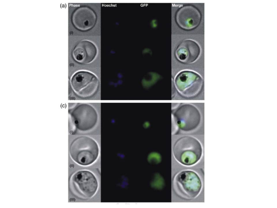
Localization of PfAspAT to the cytosol of the parasite. Localization of AspAT within P. falciparum. (a) A chimera protein consisting of the PfAspAT N-terminal region (67 aa residues) fused to GFP and (c) a fusion protein consisting of the PfAspAT C-terminal region attached to the C-terminal end of GFP were overexpressed in P. falciparum using the pARL1a expression vector. The developmental blood stages of the parasites were analyzed by fluorescent light microscopy using an Axioskop 2 plus microscope (Zeiss, Germany). (I) Ring stage; (II) trophozoite stage; (III) schizont stage. Phase, P. falciparum live image; Hoechst, staining of the parasite's nucleus; GFP, image taken using the GFP channel; Merge, merging of all images. GFP-tagged PfAspAT localizes to the cytosol in vivo.
Wrenger C, Müller IB, Schifferdecker AJ, Jain R, Jordanova R, Groves MR. Specific Inhibition of the Aspartate Aminotransferase of Plasmodium falciparum. J Mol Biol. 2010 405:956-71. Copyright Elsevier 2011