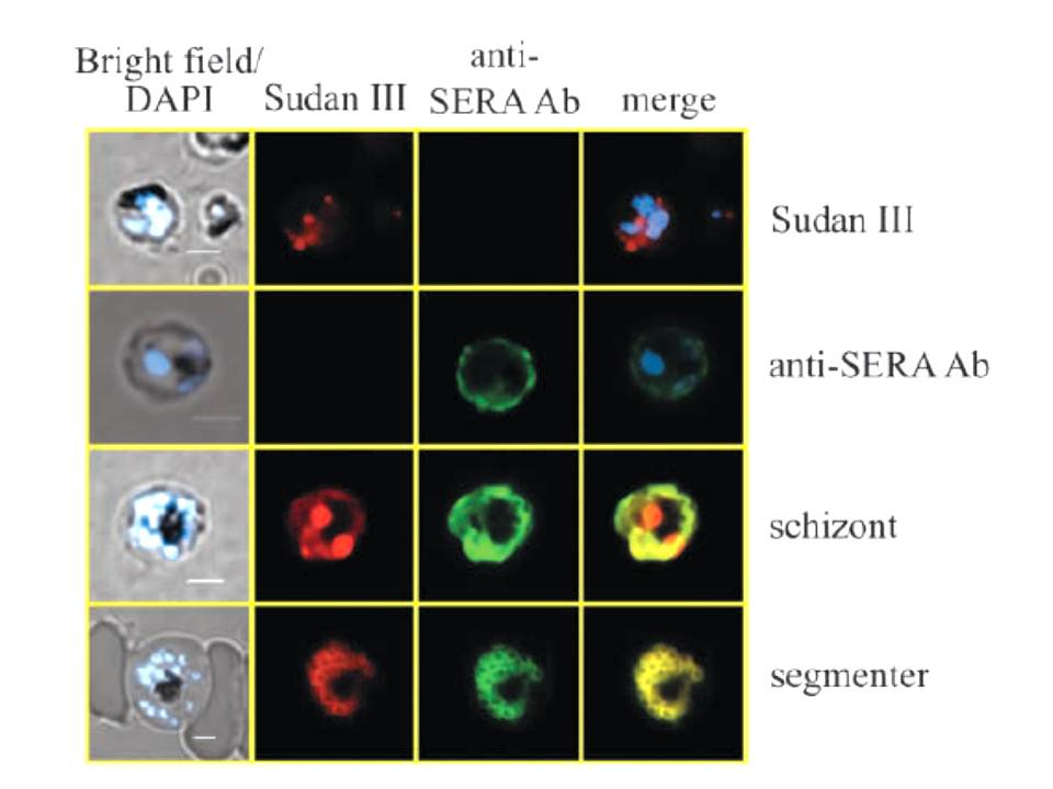
Co-localization of lipid bodies with the parasitophorous vacuole marker protein SERA. (A) Fluorescent microscopic analysis of schizont-stage P. falciparum-infected erythrocyte stained with Sudan III and anti-SERA Ab. From left to right, panels represent bright-field overlaid with DAPI, Sudan III, SERA and overlay of Sudan III and SERA. Yellow areas denote regions of overlap. Single-stained cells (top two rows) exhibit virtually no fluorescence with the opposing filter.
Palacpac NM, Hiramine Y, Mi-ichi F, Torii M, Kita K, Hiramatsu R, Horii T, Mitamura T. Developmental-stage-specific triacylglycerol biosynthesis, degradation and trafficking as lipid bodies in Plasmodium falciparum-infected erythrocytes. J Cell Sci. 2004 117:1469-80.