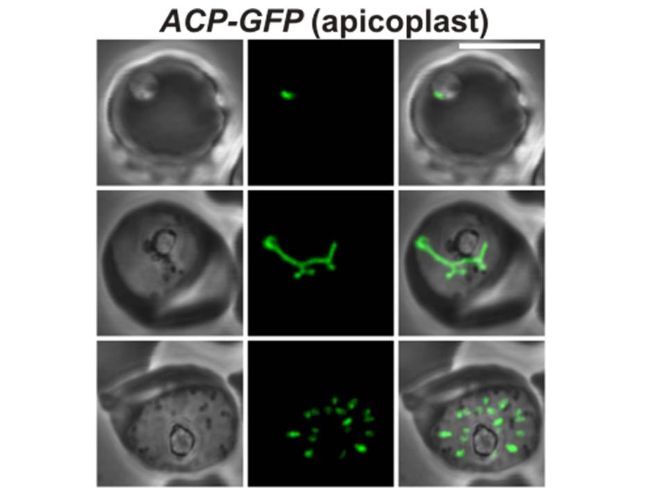
Confocal fluorescence microscopy images of transfected 3D7 P. falciparum-infected RBCs expressing a GFP chimera directed to the apicoplast. DIC image, the GFP fluorescence signal and an overlay of a P. falciparum acyl carrier protein-GFP transfectant. Scale bar = 5 µm. Apicoplasts are observed as small, rounded organelles (top row) in the ring stages that become elongated in the trophozoite stages (middle row) and forms contact points with the branched mitochondrion. The bottom row represents a schizont, where apicoplast division occurs. Apicoplast division occurs before mitochondrial division.
Tilley L, McFadden G, Cowman A, Klonis N. Illuminating Plasmodium falciparum-infected red blood cells. Trends Parasitol. 2007 23:268-77. Copyright Elsevier 2009.