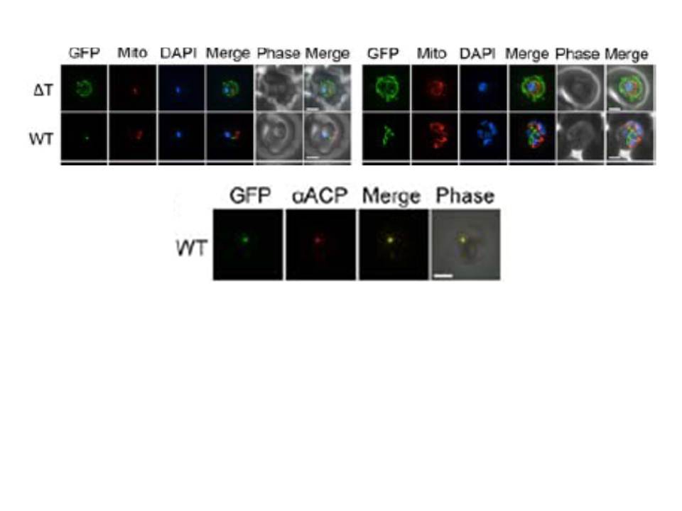
Upper panel: Epifluorescent images of live P. falciparum erythrocytic stage parasites. The parasites were stained with mitotracker to identify the mitochondrion and DAPI to identify the nucleus. GFP fluorescence in the ΔT construct (ΔT’, was generated by mimicking a previously described construct in which the TP was deleted) is associated with the parasite periphery and compartments in the erythrocyte cytosol. For the wild type (WT) construct in ring and early trophozoite stage parasites show point-like GFP fluorescence as expected for apicoplast localization. Late trophozoite stage parasites show a branched structure labeled by GFP which is distinct from the mitochondria, consistent with apicoplast localization. Image Z-stacks were deconvoluted and then presented as a single combined image. Scale bar = 2μm.
Lower panel: Colocalization of GFP constructs with endogenous ACP. αACP antibodies were affinity purified with the mature ACP domain to avoid cross-reactivity with the TP of ACP used in GFP. GFP fluorescence was found to colocalize with αACP antibodies, demonstrating apicoplast localization. Scale bar = 2μm.
Gallagher JR, Matthews KA, Prigge ST. P. falciparum apicoplast transit peptides are unstructured in vitro and during apicoplast import. Traffic. 2011 12(9):1124-38