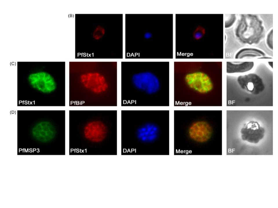
(B–D) Immunofluorescence microscopy of fixed smears of P. falciparum parasites using anti-PfStx1 antiserum. Secondary antisera (Molecular Probes) labeled with a fluorophore were used for visualization on an Olympus BX60 fluorescence microscope, and images were merged with Adobe Photoshop 5.0. In trophozoite stage parasites PfStx1 (red) has a plasma membrane like staining pattern and does not appear to be exported into the erythrocyte (B). In schizont stage parasites, PfStx1 (green) again has a peripheral location that is distinct from the ER marker PfBiP PFI0875w (in red) (C). PfStx1 (red) co-localizes with PfMSP3 (in green), a plasma membrane marker in schizont stage parasites (D), confirming its predicted location proximal to the plasma membrane.
Parish LA, Rayner JC. Plasmodium falciparum secretory pathway: Characterization of PfStx1, a plasma membrane Qa-SNARE. Mol Biochem Parasitol. 2009 164:153-6. Copyright Elsevier 2009. PMID:
Other associated proteins
| PFID | Formal Annotation |
|---|---|
| PF3D7_0210700 | syntaxin, Qa-SNARE family |