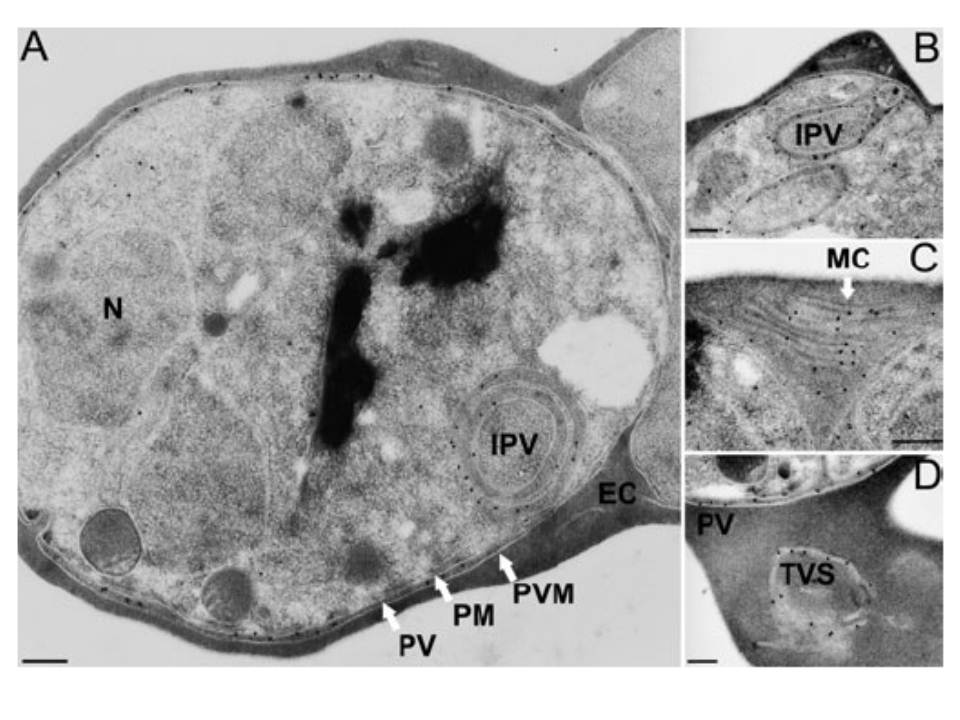
Subcellular localization of PfCDPK1. Sections of P. falciparum (clone K1) erythrocytic stages were incubated with the affinity-purified PfCDPK1-specific antibody 2129 and binding was visualized using gold-conjugated protein A. A and B. Gold particles indicative of antibody binding associated with the parasitophorous vacuole and intracellular vacuoles (intraparasitic vacuole) of schizont stages. Intracellular vacuoles originating from the plasma membrane of the parasite (B). C and D. Sections of erythrocytes infected with two schizonts and one schizont, respectively, showing labelling of the tubovesicular system in the host erythrocyte. The membranous structure visible in the erythrocyte cytosol in (C) is also known as Maurer’s clefts. EC, erythrocyte cytosol; MC, Maurer’s clefts; IPV, intraparasitic vacuole; N, nucleus; PM, parasite plasma membrane; PV, parasitophorous vacuole; PVM, parasitophorous vacuole membrane; TVS, tubovesicular system. Size bars (200 nm) have been inserted in each panel.
Möskes C, Burghaus PA, Wernli B, Sauder U, Dürrenberger M, Kappes B. Export of Plasmodium falciparum calcium-dependent protein kinase 1 to the parasitophorous vacuole is dependent on three N-terminal membrane anchor motifs. Mol Microbiol. 2004 54:676-691. Copyright John Wiley & Sons Ltd. 2010.