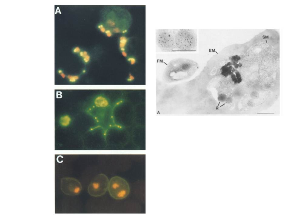
Left row: mAb 1B9 Immunofluorescence of merozoite invasion. Thin smears were incubated with mAb IB9, FITC-goat anti-mouse IgG and counterstained with ethidium bromide. (A and C) to identify parasite nucleus. (A) Merozoites attached to erythrocytes. Erythrocyte at upper right has been invaded by two merozoites. (B) Merozoites in the process of invading; rhoptry protein is discharged around erythrocyte surface. (C) Erythrocytes invaded by merozoites. Rhoptry protein is localized in infected erythrocyte membrane.
Right picture: Immunoelectron microscopic localization of the rhoptry protein using mAb 1B9 and protein A-gold. (A) post-embedded immunolabeling of mature schizont-infected erythrocyte showing localization of 5-nm gold particles over rhoptries. EM, Erythrocyte membrane; SM, schizont membrane; FM, free merozoite; R, rhoptries. (Inset) High magnification of paired rhoptry organelles showing localizaltion of rhoptry antigen.
Sam-Yellowe TY, Shio H, Perkins ME. Secretion of Plasmodium falciparum rhoptry protein into the plasma membrane of host erythrocytes. J Cell Biol. 1988 1061507-13.