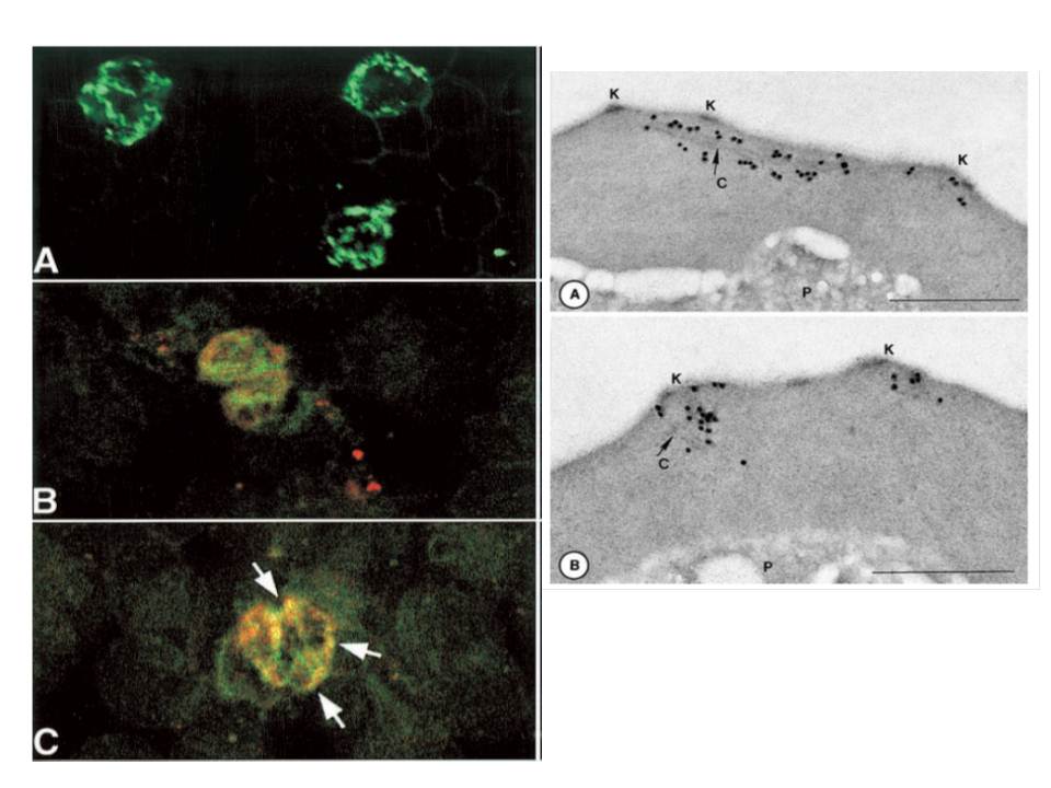
Right panel:Immunodetection of P. falciparum proteins by immuno-electron microscopy, using mAB SP1AG5. Stage-specific-infected erythrocytes were processed as described. Schizont-infected erythrocytes were reactive with antibody as shown. Gold particles (15 nm) in panel label longitudinal clefts (Maurer’s clefts, C); and panel B. They label knobs (K)
:Stage-specific IFA using mAbs. A. mAb with fluorescein
Left panelisothiocyanate-conjugated staining. B. mAb co-localize with Bodipy ceramide in trophozoites and schizonts (C). This suggests that Rhop3 localize in lipid-rich areas of the parasite, putatively within the intra-host cell cellular network.
Sam-Yellowe TY, Fujioka H, Aikawa M, Hall T, Drazba JA. A Plasmodium falciparum protein located in Maurer's clefts underneath knobs and protein localization in association with Rhop-3 and SERA in the intracellular network of infected erythrocytes. Parasitol Res. 2001 87:173-85. With kind permission of Springer Science+Business Media.