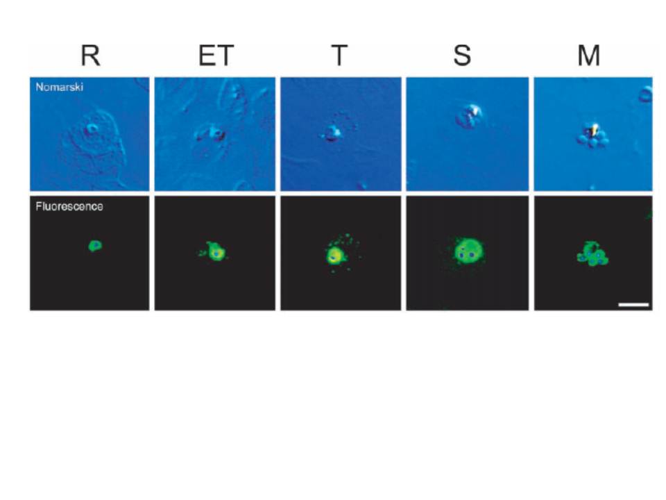
Change in the distribution of PfNSF during development. Parasitized erythrocytes at various stages as indicated were doubly labeled with antibodies against NSF and 49,6-diamidino-2-phenylindole HCl and observed under a Nomarski microscope (upper panel) and a fluorescence microscope (lower panel). After superpositioning the two images, the localization of PfNSF (green) and nuclei (blue) is shown. R, ring; ET, early trophozoite; T, trophozoite; S, schizont; M, merozoite. Bar, 5 mm. A similar degree of anti-PfNSF immunoreactivity was observed in all stages of intraerythrocyte parasites, indicating that PfNSF is expressed in all cell stages. At the early trophozoite stage, PfNSF appeared in the extraparasite space and seemed to be associated with several apparent vesicular structures outside the parasites in the erythrocyte cytoplasm. At the trophozoite stage, the PfNSF-positive vesicular structure seems to be more discrete and distributed throughout erythrocyte cytoplasm, which becomes weak at the schizont stage and disappears at the merozoite stage. These results indicate that export of PfNSF occurs at the early trophozoite stage.
Hayashi M, Taniguchi S, Ishizuka Y, Kim HS, Wataya Y, Yamamoto A, Moriyama Y. A homologue of N-ethylmaleimide-sensitive factor in the malaria parasite Plasmodium falciparum is exported and localized in vesicular structures in the cytoplasm of infected erythrocytes in the brefeldin A-sensitive pathway. J Biol Chem. 2001 276:15249-55.
