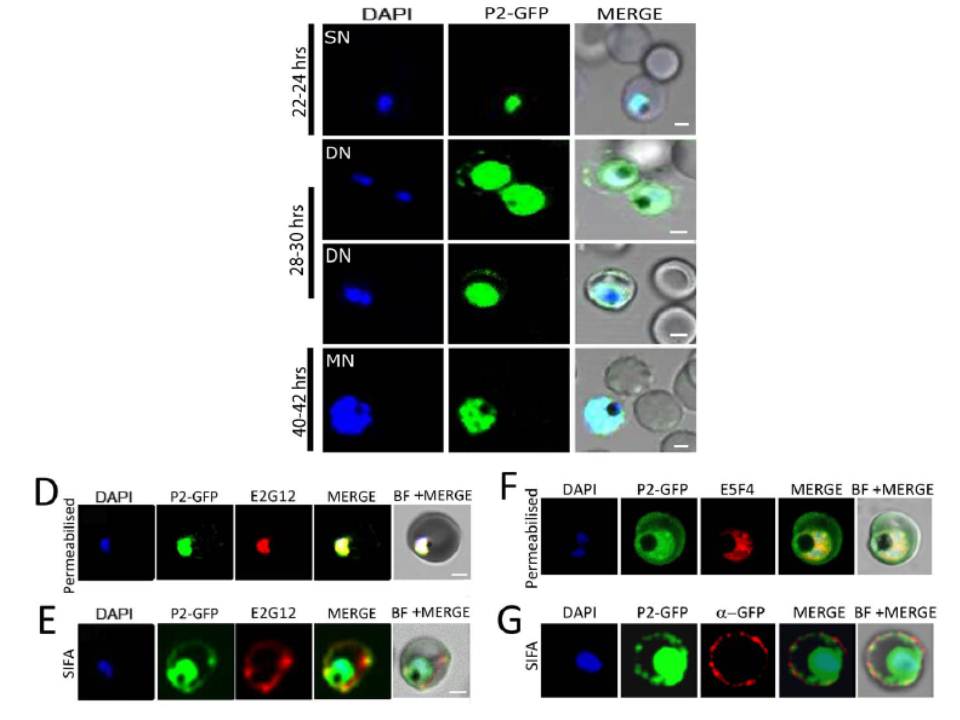
Upper panel: IFA of synchronized P. falciparum cells using DAPI (blue), P2 (green), and bright field of IE at various time points PMI in parasite development. Scale bar indicates 2 mm. Confocal microscopy of P2-GFP transfected cells at different stages of parasite growth. DAPI (blue), P2-GFP (green). GFP on the IE-surface at 28–30 hrs PMI trophozoite stage, but not at 22–24 hrs or 40–42 hrs.
Lower panel: (D,F) Confocal microscopy of permeabilized IE showing D) Red: anti-PfP2 mAb E2G12; F) Red: anti-PfP0 mAb E5F4; Green: P2-GFP staining of Pf3D7 P2-GFP infected RBCs. (E,G) Solution immunofluorescence (SIFA) of DN Pf3D7 P2-GFP infected RBCs showing E) Red: anti-PfP2 mAb E2G12; G) Red: anti-GFP antibody; Green: P2-GFP staining of Pf3D7 P2-GFP infected RBCs. Unpermeabilized SIFA at the DN stage showed that the P2 staining and GFP colocalized on the IE surface (4E).
Das S, Basu H, Korde R, Tewari R, Sharma S. Arrest of Nuclear Division in Plasmodium through Blockage of Erythrocyte Surface Exposed Ribosomal Protein P2. PLoS Pathog. 2012 Aug;8(8):e1002858.