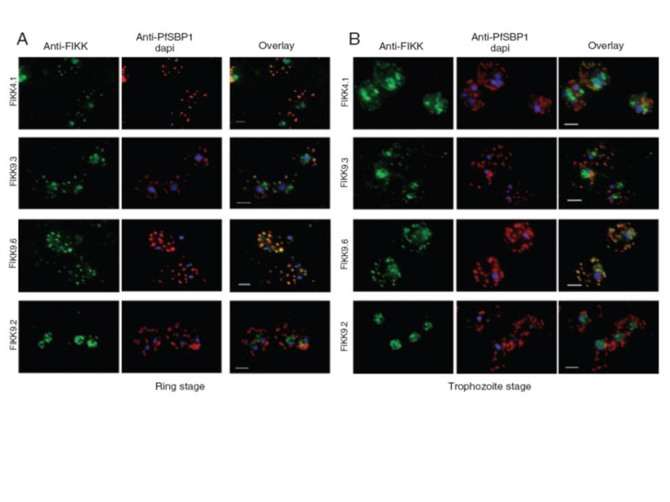
Localization by immunofluorescence of FIKK4.1, FIKK9.2, FIKK9.3 and FIKK9.6 proteins in 3D7 parasites. A and B. Ring stage (A) and trophozoite stage (B) P. falciparum-infected erythrocytes were air dried and immunolabelled with PfSBP1 antibody (red) and a FIKK-specific antibody (green). First row: polyclonal guinea pig anti-FIKK4.1; second row: polyclonal guinea pig anti-FIKK9.3; third row: polyclonal rat anti-FIKK9.6; last row: chicken anti-FIKK9.2. The third column in each panel represents the merging of the red and green channels. Yellow areas represent regions of colocalization seen on both parasitic stages for all the FIKK proteins except FIKK9.2. All proteins (not FIKK9.2) localize to the Maurer’s clefts.
Nunes MC, Goldring JP, Doerig C, Scherf A. A novel protein kinase family in Plasmodium falciparum is differentially transcribed and secreted to various cellular compartments of the host cell. Mol Microbiol. 2007 63:391-403.
Other associated proteins
| PFID | Formal Annotation |
|---|---|
| PF3D7_0424500 | serine/threonine protein kinase, FIKK family |
| PF3D7_0902100 | serine/threonine protein kinase, FIKK family |
| PF3D7_0902200 | serine/threonine protein kinase, FIKK family |