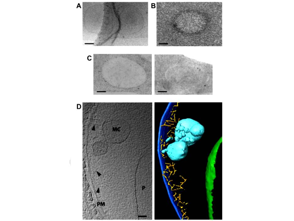
Morphology of young Maurer’s clefts in ring-infected HbAA erythrocytes. Representative electron-microscopic images of parasitized erythrocytes preserved by: A. cryo-preservation; B. high pressure freezing and freeze-substitution. Post staining with uranyl acetate revealed a densely packed protein coat surrounding the membrane of the Maurer’s cleft. C. Immuno-electron micrograph of cells preserved by high pressure freezing and freeze substitution, and immuno-labelled with an antiserum to PfSBP1 coupled to 10 nm protein A colloidal gold. D. Section through a cyro-electron tomogram (left) and corresponding surface rendered view (right) of a Maurer’s cleft precursor in a ring-infected erythrocyte. Labeling and color code as following: PM, erythrocyte plasma membrane (dark blue); MC, Maurer’s cleft precursor (cyan); P, parasite (boundary of parasite, green), actin filaments (yellow) indicated by arrowheads. Scale bars, 100 nm in all images.
Kilian N, Dittmer M, Cyrklaff M, Ouermi D, Bisseye C, Simpore J, Frischknecht F, Sanchez CP, Lanzer M. Hemoglobin S and C affect the motion of Maurer's clefts in P. falciparum-infected erythrocytes. Cell Microbiol. 2013 15(7):1111-26