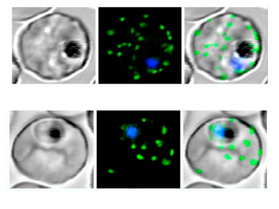
Live P. falciparum-infected erythrocytes were incubated in physiological Ringer’s solution and viewed, at 37 oC, using an LSM510 confocal laser scanning microscope. Confocal images of live P. falciparum-infected erythrocytes expressing SBP. Left images, differential interference contrast (DIC); middle images, GFP fluorescence and nuclear staining with Hoechst; right images, overlay. Scale bar, 4 μm.
The right column shows confocal live cell images of P. falciparum-infected erythrocytes expressing MAHRP. Both proteins localize to the Maurer’s clefts.
Saridaki T, Fröhlich KS, Breton CB, Lanzer M. Export of PfSBP1 to the Plasmodium falciparum Maurer's clefts. Traffic. 2008 10(2):137-52.
PubMed Article: Export of PfSBP1 to the Plasmodium falciparum Maurer's clefts
Other associated proteins
| PFID | Formal Annotation |
|---|---|
| PF3D7_1370300 | membrane associated histidine-rich protein |