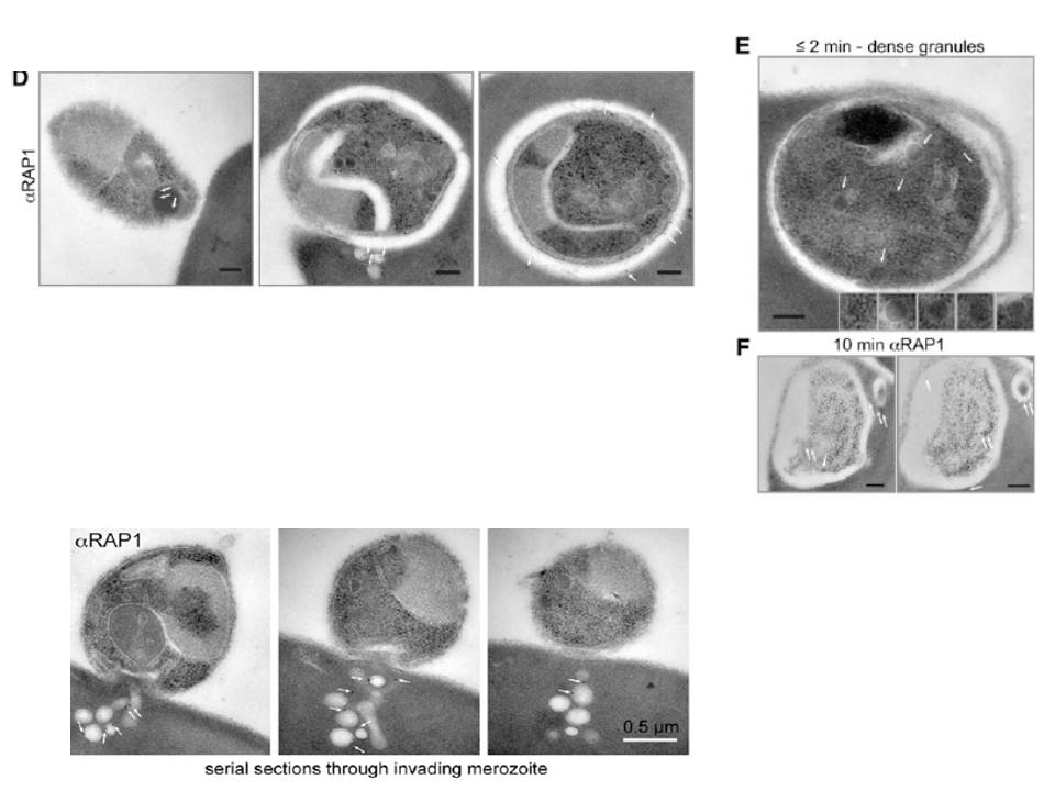
Electron microscopy with immunogold labeling of RAP1 (white arrows). The scale bar represents 0.2 mm. (E) Electron microscopy of merozoite after invasion showing intact dense granules (white arrows and insets). The scale bar represents 0.2 mm. (F) Serial sections of electron microscopy with immunogold labeling of RAP1 10 min after invasion (white arrows). The scale bar represents 0.2 mm. RAP1 labeling revealed prominent extensions of the PV into the erythrocyte cytosol.
Lower panel: Serial section electron microscopy with immunogold labeling of RAP1 showing colocalization of RAP1 with rhoptry-derived membrane into erythrocyte cytosol during an untreated merozoite invasion event.
Riglar DT, Richard D, Wilson DW, Boyle MJ, Dekiwadia C, Turnbull L, Angrisano F, Marapana DS, Rogers KL, Whitchurch CB, Beeson JG, Cowman AF, Ralph SA, Baum J. Super-resolution dissection of coordinated events during malaria parasite invasion of the human erythrocyte. Cell Host Microbe. 2011 9:9-20.