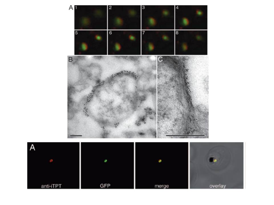
Immunolocalization of PfoTPT to the outside of free, intact apicoplasts. (A) Z-stack of immunofluorescent images (1– 8) showing localization of anti-PfoTPT (red) around apicoplast-targeted GPF (green) in parasites expressing PfACP(leader)-GFP. (B) Electron micrograph of intact apicoplast labeled with anti-PfoTPT antibodies and 20-nm colloidal gold. (C) Tannic acid fixation showing four bounding membranes around isolated apicoplast labeled with anti-PfoTPT antibodies and 10-nm colloidal gold. (Scale bars, 200 nm.)
Lowest row: Anti-PfiTPT (red) colocalizes with apicoplast-targeted GPF (green) in parasites expressing PfACP(leader)-GFP.
Mullin KA, Lim L, Ralph SA, Spurck TP, Handman E, McFadden GI. Membrane transporters in the relict plastid of malaria parasites. Proc Natl Acad Sci U S A. 2006 103:9572-7. Copyright 2009 National Academy of Sciences, U.S.A.
Other associated proteins
| PFID | Formal Annotation |
|---|---|
| PF3D7_0530200 | phosphoenolpyruvate/phosphate translocator, PPT |