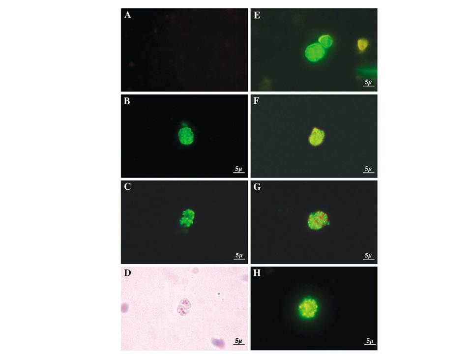
Different PfRabs have different sub-cellular distributions in schizont-infected erythrocytes. Panel (A) shows control rabbit pre-immune serum. Panel (B) shows a multinucleated schizont decorated with anti-PfRab1A antibodies giving a perinuclear staining. Panels (C) and (D) (phase contrast) show the pattern obtained with the anti-PfRab11A antibodies. Clearly, PfRab1A and PfRab11A display different distributions. Panel (E) shows the pattern obtained with anti-Pf39 (ERC) antibodies specific for the endoplasmic reticulum. Panel (F) shows a double-labelling with anti-PfRab1A (green) and Pf39 (red) antibodies, where colocalization stains yellow. Panel (G) shows a double-labelling with anti-PfRab11A (green) and anti-Pf39 (red) and no co-localization is observed. Panel (H) shows anti-PfRab6 (green) and anti-Pf39 (red), where co-localization stains yellow.
Quevillon E, Spielmann T, Brahimi K, Chattopadhyay D, Yeramian E, Langsley G. The Plasmodium falciparum family of Rab GTPases. Gene. 2003 306:13-25.
Other associated proteins
| PFID | Formal Annotation |
|---|---|
| PF3D7_0513800 | ras-related protein Rab-1A |
| PF3D7_1108600 | endoplasmic reticulum-resident calcium binding protein |
| PF3D7_1320600 | ras-related protein Rab-11A |