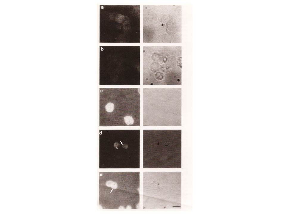
Localization of ATPase 1 by IFA. Abs to two regions of derived amino acid sequence from ATPase 1(anti-pep1 and anti-pep2) were applied to slides from isolate T9/96. Figures show dark filed (fluorescence) and bright filed views. Hemozoin pigment in bright field views corresponds to the location of parasitized red blood cells. a) Negative control (no FITC-conjugated Abs). b) negative control (no primary Abs). c) anti-Band 3 mAbs; d) anti pep1 Abs. e) anti-pep2 Abs. The arrows indicate fluorescence in the region of the parasite plasma membrane/parasitophorous vacuole, and the arrowhead indicates staining internal to the parasite. Bar: 10 mm.
Krishna S, Cowan G, Meade JC, Wells RA, Stringer JR, Robson KJ. A family of cation ATPase-like molecules from Plasmodium falciparum. J Cell Biol. 1993 120:385-98.