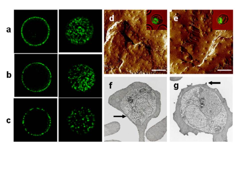
Distributions of PfEMP-1 signals and knobs on the surface of parasitized AA and CB RBCs. a–c, Confocal cross-sections through the mid-plane (left column) or maximum intensity projections of z stacks through the upper cell surface (right column) of trophozoite infected RBCs probed with a polyclonal antiserum against PfEMP-1 (FVO line). a, PfEMP-1 fluorescence patterns typical of a parasitized AA RBC. b, Fluorescence patterns from a parasitized CB RBC, similar to those of parasitized AA RBCs. c, Irregular, patchy PfEMP-1 distribution on a parasitized CB RBC. d,e, Atomic force micrographs of CB RBCs infected with the FVO P. falciparum line, showing normal AA-like (d) or abnormal (e) knob appearances. The mean (6 SEM) width of 15 randomly selected knobs in panels d and e were 50.3 nm (63.4 nm) and 96.4 nm (66.7 nm), respectively. Scale bar = 1 mm. Transmission electron micrographs of CB RBCs infected with the FVO P. falciparum line also showed populations with normal (f) or abnormal (g) knob appearances. Comparison atomic force and transmission electron micrographs of parasitized HbC and HbS RBCs can be found in references 9, 10, and 11. Arrows indicate examples of normal (f) and abnormal (g) knobs.
Amaratunga C, Lopera-Mesa TM, Brittain NJ, Cholera R, Arie T, Fujioka H, Keefer JR, Fairhurst RM. A Role for fetal hemoglobin and maternal immune IgG in infant resistance to Plasmodium falciparum malaria. PLoS One. 2011 Apr 12;6(4):e14798.