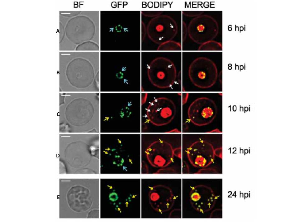
Live cell imaging showing intermediate compartments and timing of export of PfEMP1B-GFP. PfEMP1B-GFP transfectants were co-labeled with BODIPY-ceramide and samples were collected from a tightly synchronized culture (2 hours). (A) BODIPY-ceramide-labeled features (white arrows) are present in the RBC cytoplasm by 6 h post-invasion, while PfEMP1B-GFP is still located in a "necklace of beads" pattern at the parasite surface (aqua arrows). (C) At 10-12 h postinvasion PfEMP1B-GFP is observed in compartments in the RBC cytoplasm (yellow arrows). (E) By ~24 h post-invasion the PfEMP1B-GFP-labeled structures have become immobilized. Scale bars = 3 μm.
McMillan PJ, Millet C, Batinovic S, Maiorca M, Hanssen E, Kenny S, Muhle RA, Melcher M, Fidock DA, Smith JD, Dixon MW, Tilley L. Spatial and temporal mapping of the PfEMP1 export pathway in Plasmodium falciparum. Cell Microbiol. 2013 15(8):1401-18