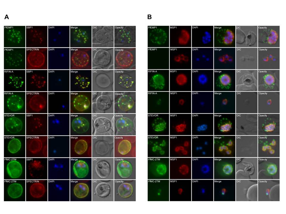
Co-localization of variant surface antigens (VSA) during the intraerythrocytic developmental cycle. A, B: Co-localization of PfEMP1, RIFIN, STEVOR and PfMC-2TM (green) with marker proteins for the erythrocyte membrane (spectrin), the Maurer’s clefts (SBP1) and the merozoite surrounding membrane (MSP1) (red). Subcellular VSA localization was determined in trophozoites (A) as well as in schizonts and free merozites (B) from clinical isolates as well as strain 3D7. Nuclei were stained with DAPI (blue). In the trophozoite stage, RIFIN proteins were exported predominantly to the Maurer’s clefts and the erythrocyte membrane, whereas STEVOR and PfMC-2TM proteins seemed to localize predominantly to the IE membrane. A-type RIFINs, STEVORs and PfMC-2TMs were located in close proximity to the nuclei in dividing parasites in the clinical isolates. In strain 3D7 schizonts, PfMC-2TM family proteins exhibited two distinct localization patterns depending on the antisera used for detection. PfMC-2TM proteins appeared to associate with the parasitophorous vacuole membrane or parasite membrane when using a-PfMC-2TM-SC, while staining with a-PfMC-2TM-CT resulted in a fluorescence pattern similar to RIFIN and STEVOR.
Bachmann A, Petter M, Tilly AK, Biller L, Uliczka KA, Duffy MF, Tannich E, Bruchhaus I. Temporal Expression and Localization Patterns of Variant surface Antigens in Clinical Plasmodium falciparum Isolates during Erythrocyte Schizogony. PLoS One. 2012;7(11): e49540.
Other associated proteins
| PFID | Formal Annotation |
|---|---|
| PF3D7_0114100 | Pfmc-2TM Maurer's cleft two transmembrane protein |
| PF3D7_0501300 | skeleton-binding protein 1 |
| PF3D7_0930300 | merozoite surface protein 1 |
| PfEMP1 | PfEMP1 |
| RIFIN | RIFIN |