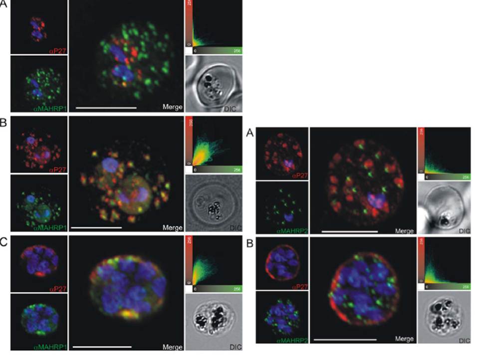
Left: Co-localization of Tex1 with MAHRP1. P27-specific polyclonal rabbit sera was used to detect Tex1 (red). Co-localization was performed using MAHRP1 polyclonal mouse sera (green). Co-localization was performed in ring stage (A) trophozoite (B) and schizont stage (C) infected RBC. Nuclear DNA was stained with DAPI (blue), Transmission image (DIC), Scale bar: 5 mm.
Right: Tex1 localization in trophozoite and schizont stages with respect to newly described structures called tethers. Colocalization of Tex1 (red) with MAHRP2 (green) A) in trophozoite stages and B) in schizont stages. Nuclear DNA was stained with DAPI (blue), Transmission image (DIC), Scale bar: 5 mm.
Kulangara C, Luedin S, Dietz O, Rusch S, Frank G, Mueller D, Moser M, Kajava AV, Corradin G, Beck HP, Felger I. Cell biological characterization of the malaria vaccine candidate trophozoite exported protein 1. PLoS One. 2012;7(10):e46112.
Other associated proteins
| PFID | Formal Annotation |
|---|---|
| PF3D7_0603400 | trophozoite exported protein 1 |
| PF3D7_1353200 | membrane associated histidine-rich protein 2 |