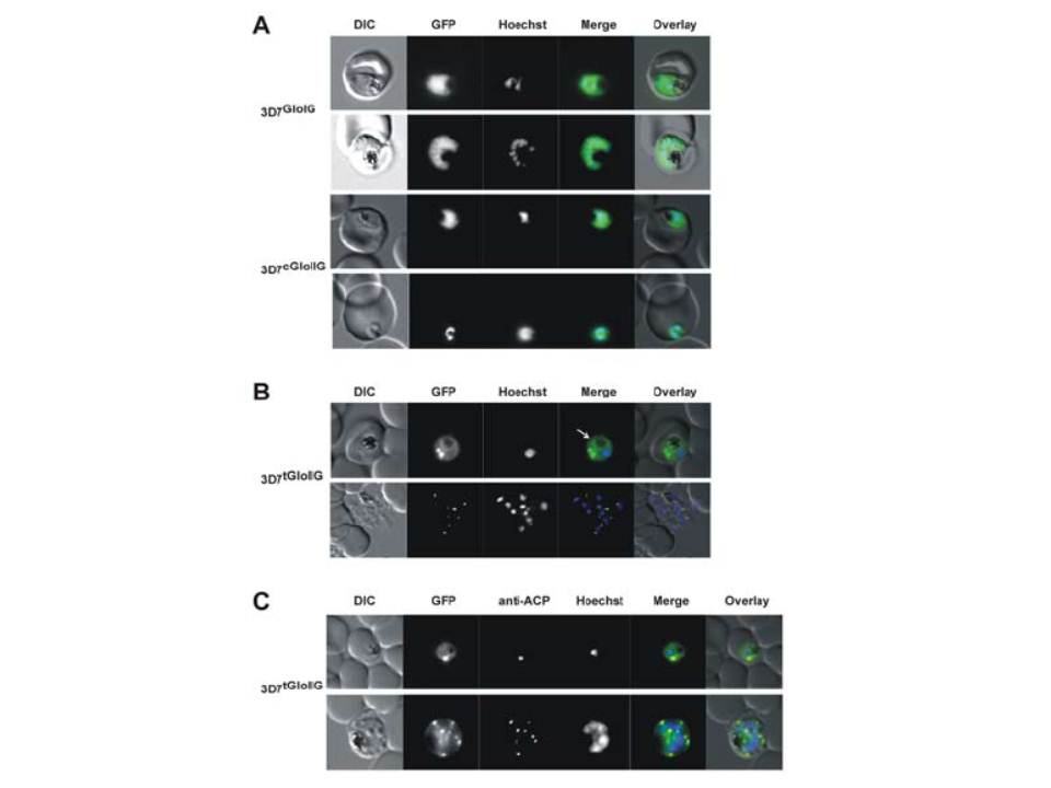
Localization of GloI, cGloII and tGloII GFP-fusion constructs in infected
erythrocytes. (A) Live cell imaging of erythrocytes infected with transgenic parasite lines 3D7GloIG (upper panels) and 3D7cGloIIG (lower panels). GFP fluorescence was evident only within the body of the parasite (excluding the food vacuole). (B) Live cell imaging of erythrocytes infected with 3D7tGloIIG (apicoplast). A distinct fluorescent “dot” was seen in both trophozoite (upper panel) and merozoite (lower panel) stage parasites, indicative of an apicoplast localization. The white arrow shows additional low levels in the arasitophorous vacuole. (C) Colocalization of tGloIIGFP and the apicoplast marker ACP in fixed, immunodecorated cells. Note the high degree of overlap between tGloII-GFP and ACP.
Urscher M, Przyborski JM, Imoto M, Deponte M. Distinct subcellular localization in the cytosol and apicoplast, unexpected dimerization and inhibition of Plasmodium falciparum glyoxalases. Mol Microbiol. 2010 76:92-103. Copyright John Wiley & Sons Ltd. 2010.
Other associated proteins
| PFID | Formal Annotation |
|---|---|
| PF3D7_0208500 | acyl carrier protein |
| PF3D7_0604700 | glyoxalase I-like protein gilp |
| PF3D7_1205700 | targeted glyoxalase II |