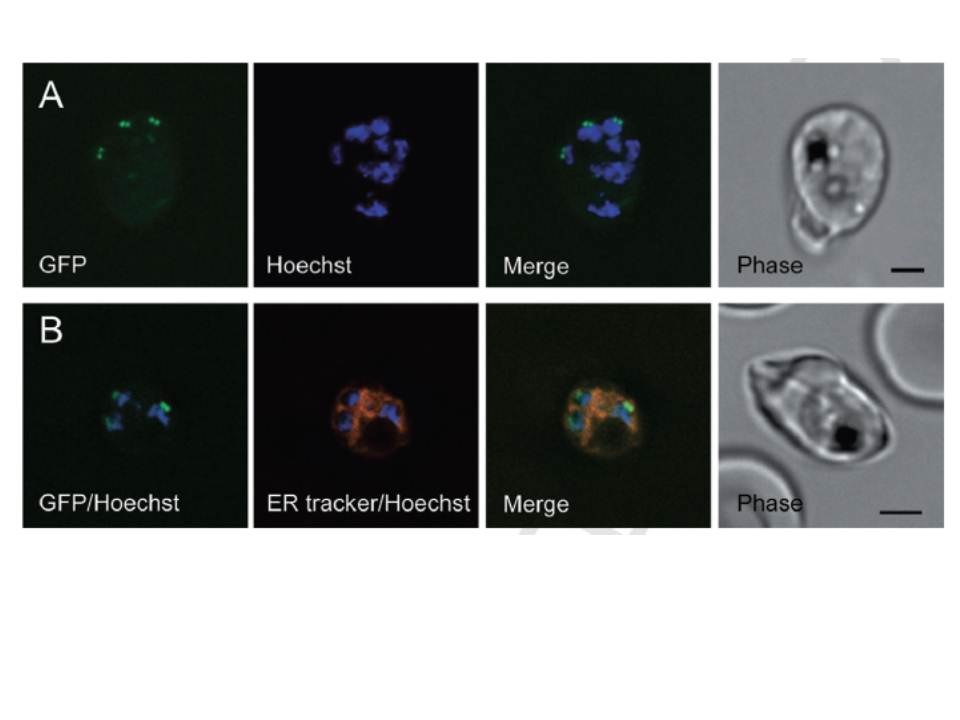
Subcellular localization of Pfark-1–GFP. Live images of schizont stage P. falciparum transgenic parasites expressing the Pfark-1–GFP protein. A. Parasites stained with Hoechst 33258. The localization of C-terminally GFP-tagged Pfark-1 protein (green signal) was observed as paired dots in close proximity to Hoechst-stained nuclear DNA (blue). B. Parasites stained with Hoechst 33258 and ER-tracker. The ER forms a highly developed membranous system from trophozoite to schizont stage. The superimposed images show that the GFP-tagged Pfark-1 protein (green dots) localize to the edge of the nuclear DNA (blue) and the ER/nuclear membrane (red), suggesting a localization of Pfark-1–GFP in contact with the nuclear envelope.
Reininger L, Wilkes JM, Bourgade H, Miranda-Saavedra D, Doerig C. An essential Aurora-related kinase transiently associates with spindle pole bodies during Plasmodium falciparum erythrocytic schizogony. Mol Microbiol. 2011 79(1):205-21.