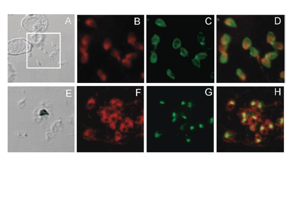
Two-color indirect immunofluorescence confocal microscopy of fixed smears of P. falciparum 3D7 merozoites. Panels A and E represent images generated by differential interference contrast (DIC) microscopy of the fixed smears incubated with (B) anti-MSP10B rabbit antiserum (red), (C) 1E1, a mAb reactive with MSP119 (green), (D) merge of micrographs B and C, (F) anti-MSP10B rabbit antiserum (red), (G) anti-RAMA mouse antiserum (green), (H) merge of micrographs F and G. The protein partitions in the detergent-enriched phase after Triton X-114 fractionation and is localized to the surfaces of trophozoites, schizonts and free merozoites in an apical distribution.
Black CG, Wang L, Wu T, Coppel RL. Apical location of a novel EGF-like domain-containing protein of Plasmodium falciparum. Mol Biochem Parasitol. 2003 127:59-68. Copyright Elsevier 2009. PMID: