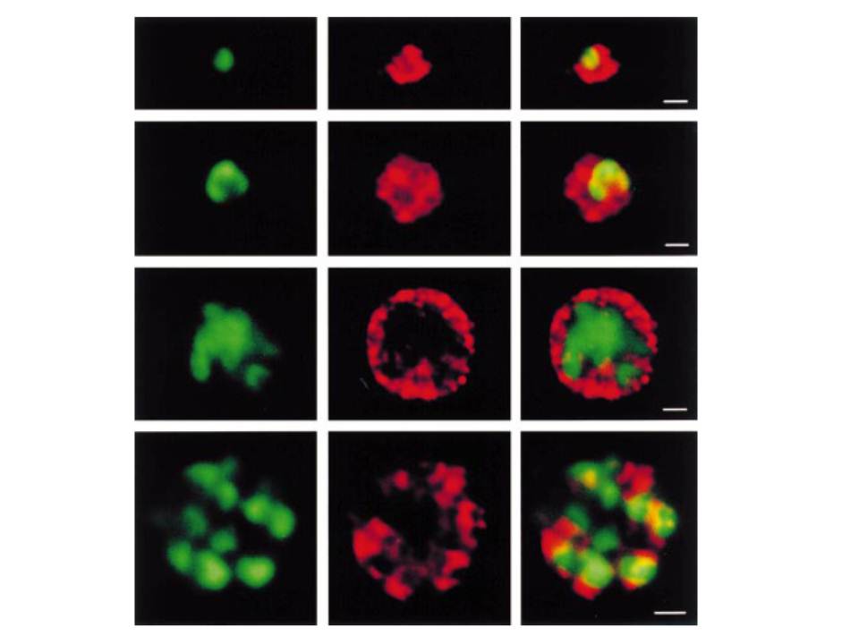
Stage-specific localization of PfCS using confocal microscopy. The rows in each column represent four parasitic stages. First row (from the top), ring stage; second row, trophozoite; third row, schizont; fourth row, segmenter. The scale bars represent 1 mm. Left column (green), DNA stain with Hoechst dye; centre column (red), PfCS immunofluorescence with AbPfCS1; right column, composite. The appearance of a dispersed immunofluorescence pattern surrounding the nuclear stain and the absence of a discrete crescent-shaped signal clearly indicate that PfCS is cytosolic and not located to the apicoplast.
Fitzpatrick T, Ricken S, Lanzer M, Amrhein N, Macheroux P, Kappes B. Subcellular localization and characterization of chorismate synthase in the apicomplexan Plasmodium falciparum. Mol Microbiol. 2001 40:65-75.