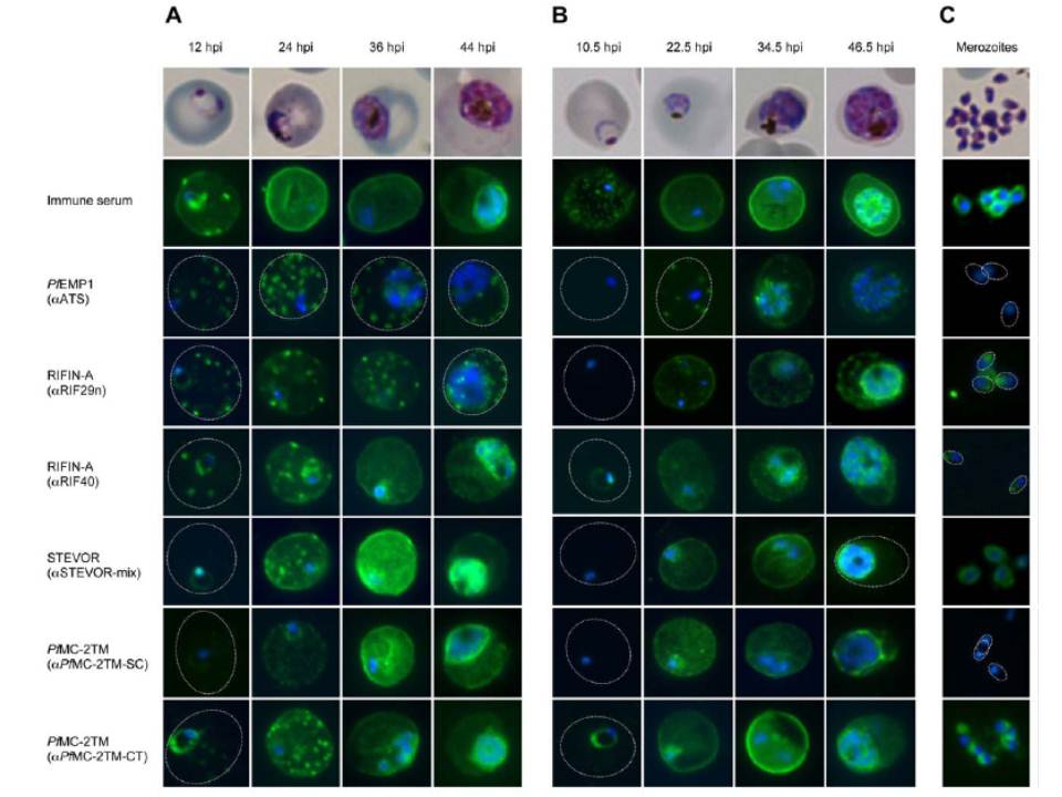
Localization of VSAs during the intraerythrocytic developmental cycle. A-C: Representative immunofluorescence images of the indicated VSAs in different parasite developmental stages of clinical isolate #4 (A), 3D7 parasites (B), and free merozoites from isolate #1 (C). First row: Giemsa staining of the corresponding parasitic stage. Second row: Positive control serum obtained from a semi-immune patient. Third row: PfEMP1-specific antibody, showing the presence of the protein in Maurer’s clefts over the entire time course (A, B). Third to eighth rows: 2TM proteins were exported into the host cell (12–36 hpi) during the trophozoite stage but remained inside the parasite in the schizont stage (48 hpi). Proteins of the RIFIN-A family frequently localized to Maurer’s clefts, particularly when using the a-RIF29n antiserum, and the erythrocyte membrane; STEVOR and PfMC-2TM localized predominantly to the erythrocyte membrane (A, B). RIFIN and STEVOR proteins were also observed at the apical tip or at the merozoite membrane, respectively. Isolate #1 also exhibited PfMC-2TM-specific fluorescence in free merozoites when using the a-PfMC-2TM-CT antiserum (C). All antibodies were visualized with Alexa488-conjugated secondary antibody (green), and nuclei were stained with DAPI (blue).
Bachmann A, Petter M, Tilly AK, Biller L, Uliczka KA, Duffy MF, Tannich E, Bruchhaus I. Temporal Expression and Localization Patterns of Variant surface Antigens in Clinical Plasmodium falciparum Isolates during Erythrocyte Schizogony. PLoS One. 2012;7(11): e49540. PMID:
Other associated proteins
| PFID | Formal Annotation |
|---|---|
| PF3D7_0631400 | Pfmc-2TM Maurer's cleft two transmembrane protein |
| PfEMP1 | PfEMP1 |
| RIFIN | RIFIN |